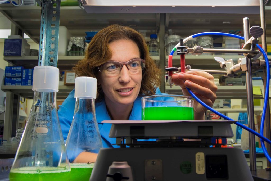
Jerilyn Timlin
Distinguished Member of the Technical Staff
Distinguished Member of the Technical Staff
(505) 844-7932
Sandia National Laboratories, New Mexico
P.O. Box 5800
Albuquerque, NM 87185-0895
Biography
and spectroscopic-based tools in conjunction with multivariate image analysis to understand the fundamental spatial-temporal relationships that govern complex biological processes. Her work interfaces analytical chemistry and optical physics with molecular and cellular biology and has direct impact on human health, plant science, and renewable energy research. She has initiated and collaborated with researchers from different scientific disciplines to tackle complex problems in biodefense and bioenergy, including multiplexed imaging of endogenous and exogenous fluorescence in plant and animal cells and tissues, visualization of host-pathogen interactions, nanoparticle uptake and trafficking, receptor ligand interactions, and identification of molecular biomarkers for early detection of disease and cellular response to changing environmental conditions.
Education
Bachelor’s Degree: Chemical Engineering, Geneva College, Beaver Falls, PA (1995)
Doctoral Degree: Analytical Chemistry, University of Michigan, Ann Arbor, MI (2000)
Postdoctoral Fellowship: Analytical Chemistry, Sandia National Laboratories (2002)
Research Interests
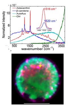
Timlin’s research focuses on developing and applying novel analytical-imaging and multivariate-analysis tools to elucidate complex spatial-temporal relationships of a variety of biomolecules that drive key biological processes.The work in our lab crosses traditional boundaries of chemistry, physics, and biology and often covers multiple spatial scales from single molecules on up to single cells, communities, and tissues. A main component of our work is spectral imaging – a method whereby spectrally resolved information is obtained at every two-dimensional pixel or three dimensional voxel. I have been developing and applying various spectral imaging technologies (also called chemical imaging) since my graduate work which pioneered the use of hyperspectral Raman microscopy with near-infrared excitation to elucidate the dynamic chemical composition of bone without the use of labels. Most recently, my group has employed both hyperspectral Raman and fluorescence microscopy to a variety of applications looking at single molecules to intact tissue, including the visualization of subcellular pigment distribution in photosynthetic organisms such as cyanobacteria and green algae (Figure 1 at right).
The addition of a spectral dimension can result in a three-, four-, or five-dimension image data set (2-3 spatial, 1 spectral, and 1 temporal) that is beyond human visualization capabilities. For this reason we utilize sophisticated multivariate analysis tools to mathematically extract the underlying spectral signatures and create quantitative spatial-temporal profiles of biomolecules.
In keeping with the multicolor theme, Timlin’s lab has developed multi-color, optical super resolution capabilities. Building from our unique capabilities in dual-color, video rate, total internal reflection fluorescence (TIRF) microscopy, we have constructed a simultaneous, dual-color single molecule, stochastic optical super-resolution (STORM) microscope. With spatial resolutions approaching those of electron microscopy (~30-40 nm), this technology is opening our eyes to a variety of biological processes never before seen. For example, Figure 2 highlights receptor reorganization at the membrane during immune response.
Timlin’s work interfaces analytical chemistry and optical physics with molecular and cellular biology and has direct impact on human health, plant science, and renewable energy research. We work collaboratively with researchers from different scientific disciplines to tackle complex bioscience problems.
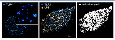
Figure 1 (top right): Hyperspectral confocal Raman image of carotenoids and chlorophyll in living Haematococcus pluvialis cells. Upper panel: component spectra. Lower panel: pigment localization, pseudo-colored corresponding to spectra in upper panel. Figure generated by Aaron Collins in collaboration with Thomas Beechem, and Howland Jones at SNL and Dr. Qiang Hu’s group at ASU.
Figure 2 (bottom left): Nanoscale organization of Toll-like Receptor 4 (TLR4) and E. coli lipopolysaccharide (LPS) at the plasma membrane of mouse macrophage cells as visualized with simultaneous, dual-color stochastic optical reconstruction microscopy (STORM). Figure generated by Jesse Aaron, work in collaboration wtih Bryan Carson at SNL.
Multiplexed Measurements of Protein Dynamics and Interactions at Extreme Resolutions
This project was funded under the National Institutes of Health (NIH) Director’s New Innovator Award in 2009. When successful, this project will provide an unprecedented view of protein interactions in the living cell through the development of novel spectrally resolved superresolution microscopy methods.
Analyzing and Understanding of Transporters to Control Lignin Transformation into Fuel
The overall goal of this project is to understand the fundamental relationships that govern lignin transport in order to control transport in a way that will provide an increase in the range and kinetics of transport into cells. Timlin is developing analytical tools for assessing lignin uptake and kinetics in bulk and at the single cell/single molecule level. Combining experimental measurements with computational modeling, she hopes to identify the types of transporters that are active on lignin and their substrate specificity as well the kinetics of lignin transport in natural and engineered hosts.
Photosynthetic Antennae Research Center (PARC)
PARC is one of 46 Energy Frontier Research Centers (EFRCs) established nationally at universities, national laboratories, nonprofit organizations, and private firms by the U.S. Department of Energy’s Office of Science. A multi-institutional collaboration, PARC seeks to understand the basic scientific principles that underpin the efficient functioning of natural photosynthetic antenna systems. These principles will then be used as a basis for man-made systems to convert sunlight into fuels. Timlin’s specific work within PARC will use advanced spectral imaging and analysis methodologies to isolate fluorescent signatures from natural and bio-inspired photosynthetic pigments to increase our understanding of the spatial distribution and abundance of these critical components in the energy transfer cascade.
Awards, Honors, and Memberships
Memberships
- Editorial board, American Journal of Molecular and Cellular Biology (2011)
- Member, Optical Society of America (2011) Member, Microscopy Society of America (2007)
- Member, Coblentz Society (2001) Member, Society for Applied Spectroscopy (1998)
- Member, American Chemical Society (1992)
Awards and Honors
- SNL Distinguished Mentorship Award (2016)
- SNL Employee Recognition Award: Individual Values, Ethics, and Integrity (2013)
- Outstanding Women at Sandia National Laboratories winner (2011)
- KNME Science Café Speaker (2011)
- NIH New Innovator Award (2009)
- R&D 100 Award Winner “Hyperspectral Confocal Microscope” (2009)
- Sandia National Laboratories Emerging Leaders Program (2008)
- American Chemical Society- Div of Analytical Chemistry Fellowship (1999-2000)
- Graduate Student Council in Chemistry President (1998-1999)
- Student panelist for Office of Conflict Resolution (1997-1999)
- GAANN (Graduate Assistantship in Area of National Need) Fellow (1995-1998)
- Graduate Student Council in Chemistry (1995-1997)
- Presidential Scholar, Geneva College (1991-1995)
Selected Patents
- US Patent. No.: US 8,686,363 B1 “Hyperspectral Stimulated Emission Depletion Microscopy and Methods of Use Thereof”, Jerilyn A. Timlin and Jesse S. Aaron, Awarded April 1, 2014.
Selected Book Chapters
- Timlin, JA, Collins, AM, Shumskaya, M, Wurtzel, ET, Beechem, TA, “Localizing and Quantifying Carotenoids in Intact Cells and Tissues” in Carotenoids. Cvetkovic, D and Nikolic, G. InTech:Rijeka, 2017, Ch. 03.
- Haaland, DM, Jones, HDT and Timlin, JA, “Experimental and data analytical approaches to automating multivariate curve resolution in the analysis of hyperspectral images ” in Resolving Spectral Mixtures, Ruckebusch, C, editor(s). Elsevier: Amsterdam, 2016, 381-406.
- Anthony S, Carroll-Portillo A and Timlin JA, “Dynamics and interactions of individual proteins in the membrane of single, living cells ” in Single Cell Protein Analysis. Singh, AK and Chandrasekaran, A, editor(s). Springer New York: 2015, 185-207.
- Chen W, Han D, Li Y, Jones HDT, Timlin JA, Hu Q, Semi-Quantitative and Absolute Quantitative Analyses of Biochemical Composition of Microalgae. In Handbook of Microalgal Cultures – Second Edition. Hu R, (Ed.), Wiley-Blackwell, 2013.
- Aaron JS and Timlin JA “Advanced Optical Imaging of Endocytosis,” in Molecular Recognition of Endocytosis, B. Ceresa (Ed.), InTech, 2012.
- Timlin JA, “Scanning microarrays: Current methods and future directions,” In DNA Microarrays, Part B: Databases and Statistics. Kimmel A, Oliver B, editors. Academic Press: New York, 2006: 79-98.
Publications
-
Timlin, J.A. (2024). Hyperspectral Fluorescence and Raman Microscopy for Characterizing Plant Response at the Cellular and Subcellular Level [Presentation]. https://doi.org/10.2172/2585874 Publication ID: 152936
-
Timlin, J.A. (2024). Hyperspectral Fluorescence and Raman Microscopy for Characterizing Plant Response at the Cellular and Subcellular Level [Conference Presentation]. https://doi.org/10.2172/2585874 Publication ID: 158232
-
Butler, K., Timlin, J.A., & Lowrey, L. (2024). In Situ Control of Bio-Inorganic Bone Production and Resorption: Towards the Development of Self-Healing Materials [Conference Poster]. https://doi.org/10.2172/2586256 Publication ID: 159516
-
Hayes, D.C., McDonald, A., Pattison, K., Butler, K., Timlin, J.A., & Piontkowski, Z.T. (2024). Label-Free, Noninvasive Bone Cell Classification by Hyperspectral Confocal Raman Microscopy. Chemical and Biomedical Imaging, 2(2), pp. 147-155. https://doi.org/10.1021/cbmi.3c00106 Publication ID: 122616
-
Katinas, C.M., Finley, P.D., Hare, A., Verzi, S.J., Slater, J., Silva, A.R., Reichardt, T.A., & Timlin, J.A. (2023). Utilizing Physics-informed and Machine Learning Methods to Enhance Remote Monitoring of Physiological Signatures [Conference Presentation]. https://doi.org/10.2172/2430787 Publication ID: 126684
-
Timlin, J.A., Katinas, C.M., Slater, J., Hare, A.T., Verzi, S.J., Finley, P.D., & Reichardt, T.A. (2023). Remote Monitoring of Physiological Signatures: The Impact of Hyperspectral Imaging [Conference Poster]. https://doi.org/10.2172/2431728 Publication ID: 129932
-
Hayes, D.C., Butler, K., Pattison, K., & Timlin, J.A. (2022). Synthetic Biology Enabled Modification of Bone Cells and the Impact on Bone Mineral Formation [Conference Poster]. https://www.osti.gov/biblio/2005723 Publication ID: 120028
-
Lane, T., Timlin, J.A., Reichardt, T.A., Fisher, C., Reese, K., Moorman, M., & Lane, P. (2022). Towards integrated pest management for algal production systems [Conference Presentation]. https://doi.org/10.2172/2005855 Publication ID: 120172
-
Katinas, C.M., Timlin, J.A., Slater, J., & Reichardt, T.A. (2022). Remote Detection Of Human Vital Signs Via Hyperspectral And Multispectral Images [Conference Poster]. https://doi.org/10.2172/2005877 Publication ID: 120260
-
Timlin, J.A., Katinas, C.M., Slater, J., Verzi, S.J., Finley, P.D., Silva, A.R., & Reichardt, T.A. (2022). Multi-modal Imaging for Remote Detection of Human Vital Signs: Towards Long Range Monitoring [Conference Presentation]. https://doi.org/10.2172/2005891 Publication ID: 120316
-
Hayes, D.C., Butler, K., & Timlin, J.A. (2022). Synthetic Biology Enabled Modification of Bone Cells and the Impact on Bone Mineral Formation [Conference Poster]. https://doi.org/10.2172/2004532 Publication ID: 117316
-
Katinas, C.M., Timlin, J.A., Slater, J., & Reichardt, T.A. (2022). Hyperspectral Signature Analysis and Characterization in Support of Remote Detection of Chemical and Biological Exposures [Conference Presentation]. Proceedings of SPIE – The International Society for Optical Engineering. https://doi.org/10.2172/2001758 Publication ID: 108116
-
Katinas, C.M., Timlin, J.A., Slater, J., & Reichardt, T.A. (2021). Hyperspectral Signature Analysis and Characterization in Support of Remote Detection of Chemical and Biological Exposures [Conference Paper]. https://doi.org/10.1117/12.2618425 Publication ID: 107760
-
Maes, D., Finke, A., Smallwood, C.R., Timlin, J.A., Ruffing, A.M., & Howard, M. (2021). Hyperspectral Bioindicators of Heavy Metal Exposure in Tall Fescue [Conference Poster]. https://doi.org/10.2172/1884053 Publication ID: 79424
-
Timlin, J.A., Reichardt, T.A., Maes, D., Kunstadt, C.G., Katinas, C.M., Hipple, T., Lane, T., Jensen, T., Dempster, T.A., McGowen, J.A., Poorey, K., & Curtis, D.J. (2021). Real-time monitoring of algal pond productivity and pest presence [Conference Presentation]. https://doi.org/10.2172/1873286 Publication ID: 78889
-
Timlin, J.A. (2021). Barriers to Scale: Algae Crop Protection Workshop- Spectroradiometric Monitoring [Presentation]. https://www.osti.gov/biblio/1864496 Publication ID: 78174
-
Maes, D., & Timlin, J.A. (2020). Concurrent evaluation of autophagy induction and Burkholderia infection at the single cell level [Presentation]. https://www.osti.gov/biblio/1825032 Publication ID: 71149
-
Maes, D., Timlin, J.A., Anthony, S.M., Podlevsky, J., & Branda, S.S. (2020). Concurrent evaluation of autophagy induction and Burkholderia infection at the single cell level [Presentation]. https://www.osti.gov/biblio/1831029 Publication ID: 71479
-
Reichardt, T.A., Maes, D., Jensen, T.J., Dempster, T.A., McGowen, J.A., Poorey, K., Curtis, D.J., Lane, T., & Timlin, J.A. (2020). Spectroradiometric detection of competitor diatoms and the grazer Poteriochromonas in algal cultures. Algal Research, 51. https://doi.org/10.1016/j.algal.2020.102020 Publication ID: 106264
-
Maes, D., Timlin, J.A., Anthony, S.M., Podlevsky, J., Courtney, C.M., & Branda, S.S. (2020). Concurrent evaluation of autophagy induction and Burkholderia infection at the single cell level. https://doi.org/10.2172/1668463 Publication ID: 99832
-
Reichardt, T.A., & Timlin, J.A. (2020). Spectroradiometric detection update [Presentation]. https://www.osti.gov/biblio/1820271 Publication ID: 74788
-
Hirakawa, M., Krishnakumar, R., Timlin, J.A., Carney, J., & Butler, K. (2020). Gene editing and CRISPR in the clinic: Current and future perspectives. Bioscience Reports, 40(4). https://doi.org/10.1042/bsr20200127 Publication ID: 73844
-
Khoshravesh, R., Pallikkuth, S., Timlin, J.A., Lidke, K., & Hanson, D. (2020). Hyperspectral Raman Imaging to Fingerprint Key Plants Metabolites and Cellular Component [Presentation]. https://www.osti.gov/biblio/1767899 Publication ID: 72760
-
Maes, D., Dailey, M., Cox, K.M., Anthony, S.M., Carson, B., Bradfute, S., Henderson, I., Ortner, M., & Timlin, J.A. (2019). Potential of Proprietary Molecules for Inducing Autophagy and Enhancing Antibiotic Treatment of Tuberculosis [Conference Poster]. https://www.osti.gov/biblio/1761289 Publication ID: 70763
-
Johnston, R.K., Seamon, K.J., Saada, E.A., Podlevsky, J., Branda, S.S., Timlin, J.A., & Harper, J.C. (2019). Use of anti-CRISPR protein AcrIIA4 as a capture ligand for CRISPR/Cas9 detection. Biosensors and Bioelectronics, 141(C). https://doi.org/10.1016/j.bios.2019.111361 Publication ID: 70297
-
Harmon, B.N., Timlin, J.A., & Ting, C. (2019). Genomic Security Related Projects [Presentation]. https://www.osti.gov/biblio/1645776 Publication ID: 70491
-
Li, K., & Timlin, J.A. (2019). Examining the Potency of Potential Autophagy Inducing Drugs [Presentation]. https://www.osti.gov/biblio/1807082 Publication ID: 69715
-
Hipple, T., Timlin, J.A., & Johnston, R.H. (2019). Quantifying protein loading in individual mesoporous silica nanoparticles [Presentation]. https://www.osti.gov/biblio/1807066 Publication ID: 69723
-
Caswell, J., Ting, C., Cuellar, C.R., Bisila, J.D., Ho, M., Wilson, B., Cairns, K., & Timlin, J.A. (2019). Automated Recognition of Dual Use Publications [Conference Poster]. https://www.osti.gov/biblio/1640779 Publication ID: 69154
-
Ting, C., Caswell, J., Cuellar, C.R., Bisila, J.D., Ho, M., Wilson, B., Cairns, K., & Timlin, J.A. (2019). Automated Recognition of Dual Use Publications [Conference Poster]. https://www.osti.gov/biblio/1640780 Publication ID: 69155
-
Lane, T., Timlin, J.A., Reichardt, T.A., Poorey, K., Lane, P., & Hagerstrand, A. (2019). A survey of microalgal resistance to algal pond crashes [Conference Poster]. https://www.osti.gov/biblio/1640882 Publication ID: 69297
-
Johnston, R.H., Saada, E.A., Podlevsky, J., Branda, S.S., Harper, J.C., & Timlin, J.A. (2019). Anti-CRISPR Based Platform for Rapid Detection and Quantification of Cas9-RNP [Conference Poster]. https://www.osti.gov/biblio/1640979 Publication ID: 69447
-
Ye, D., Hayes, D.C., Maes, D., Simoes, C.T., Appelhans, L.N., Kent, M.S., & Timlin, J.A. (2019). Transport of lignin-beakdown products into bacteria and fungi [Conference Poster]. https://www.osti.gov/biblio/1639882 Publication ID: 68303
-
Timlin, J.A. (2019). Direct Measurement of Uptake and Intracellular Accumulation of Lignin Breakdown Products in Fungi and Bacteria [Presentation]. https://www.osti.gov/biblio/1599544 Publication ID: 66064
-
Maes, D., Dailey, M., Anthony, S.M., Carson, B., Bradfute, S., Henderson, I., Ortner, M., & Timlin, J.A. (2019). Potential for Synergistic Effect of Proprietary Autophagy Inducing Molecules in the Treatment of Tuberculosis [Conference Poster]. https://www.osti.gov/biblio/1601100 Publication ID: 66988
-
Anthony, S.M., Miller, P.R., Timlin, J.A., & Polsky, R. (2019). Imaging effectiveness calculator for non-design microscope samples. Applied Optics, 58(22), pp. 6027-6037. https://doi.org/10.1364/AO.58.006027 Publication ID: 65323
-
Timlin, J.A., Johnston, R.K., Koh, C.Y., Harper, J.C., Seamon, K., Phaneuf, C., Harmon, B.N., Eckles, T.P., Saada, E.A., & Meagher, R.J. (2018). Development of Rapid Diagnostic Tools for Detection and Quantification of Cas9 Presence and Activity [Conference Poster]. https://www.osti.gov/biblio/1807018 Publication ID: 60458
-
Kuether, R.J., Allensworth, B.M., Backer, A., Chen, E.Y., Dingreville, R.P.M., Forrest, E.C., Knepper, R.A., Tappan, A.S., Marquez, M.P., Vasiliauskas, J.G., Rupper, S., Grant, M.J., Atencio, L.C., Hipple, T., Maes, D., Timlin, J.A., Ma, T.J., Garcia, R.J., Danford, F.L., … Swiler, L.P. (2018). Posters for AA/CE Reception [Presentation]. https://www.osti.gov/biblio/1592652 Publication ID: 59884
-
Timlin, J.A., & Reichardt, T.A. (2018). Remote Sensing of Algal Growth in Waterways [Presentation]. https://www.osti.gov/biblio/1592259 Publication ID: 59077
-
Harper, J.C., Johnston, R.K., Seamon, K.J., Saada, E.A., Branda, S.S., & Timlin, J.A. (2018). Rapid Electrochemical Detection of CRISPR/Cas9 Components using anti-CRISPR Protein AcrIIA4 as Capture Ligand [Conference Poster]. https://www.osti.gov/biblio/1581727 Publication ID: 63690
-
Johnston, R.K., Seamon, K.J., Saada, E.A., Podlevsky, J., Branda, S.S., Harper, J.C., & Timlin, J.A. (2018). Rapid Electrochemical Detection and Quantification of CRISPR/Cas9 Components [Conference Poster]. https://www.osti.gov/biblio/1582212 Publication ID: 63819
-
Hipple, T., & Timlin, J.A. (2018). Effects of Vampirovibro Chorellavorus on Chlorella Sorokiniana DOE Strain 1044 [Presentation]. https://www.osti.gov/biblio/1568998 Publication ID: 62998
-
Atencio, L.C., & Timlin, J.A. (2018). Effects of Vampirovibro chlorellavorus on Chlorella sorokiniana DOE 1116 [Presentation]. https://www.osti.gov/biblio/1569431 Publication ID: 63032
-
Timlin, J.A., & Quintana, V. (2018). Super-Resolution Imaging of Murine Macrophage Cells [Presentation]. https://www.osti.gov/biblio/1806640 Publication ID: 63285
-
Maes, D., Reichardt, T.A., Jensen, T.J., Dempster, T.A., McGowen, J.A., Poorey, K., Hipple, T., Lane, T., & Timlin, J.A. (2018). Spectroradiometric Detection of Competitors and Predators in Algal Ponds [Conference Poster]. https://www.osti.gov/biblio/1526346 Publication ID: 62531
-
Dailey, M., Zhang, Y., Zhang, R., Miller, E.S., Williams, P.G., Lindsey, J.S., & Timlin, J.A. (2018). Identification of the localization of tolyporphins unusual tetrapyrroles in a microbial photosynthetic community [Conference Poster]. https://www.osti.gov/biblio/1529471 Publication ID: 62647
-
Hudson, C.M., Williams, K.P., & Timlin, J.A. (2018). CasAnn: Targeted Identification of CRISPR Associated Gene Operons from Next Generation Sequences [Conference Poster]. https://www.osti.gov/biblio/1529773 Publication ID: 62727
-
Johnston, R.K., Harper, J.C., & Timlin, J.A. (2018). Development of an Antibody Based Electrochemical Platform for Cas9 Detection [Conference Poster]. https://www.osti.gov/biblio/1502283 Publication ID: 61138
-
Dailey, M., Ye, D., Sale, K.L., Maes, D., Simoes, C.T., Appelhans, L.N., Hayes, D.C., Kent, M.S., & Timlin, J.A. (2018). Accumulation of lignin-breakdown products in several lignolytic and non-lignolytic bacteria and fungi [Conference Poster]. https://www.osti.gov/biblio/1495744 Publication ID: 60689
-
Lino, C.A., Harper, J.C., Carney, J., & Timlin, J.A. (2018). Delivering crispr: A review of the challenges and approaches. Drug Delivery, 25(1), pp. 1234-1257. https://doi.org/10.1080/10717544.2018.1474964 Publication ID: 58931
-
Hudson, C.M., Timlin, J.A., & Williams, K.P. (2017). CasANN: Targeted Identification of CRISPR Associated Gene Operons from Next Generation Sequences [Conference Poster]. https://www.osti.gov/biblio/1513657 Publication ID: 58783
-
Dailey, M., Ye, D., Maes, D., Simoes, C.T., Appelhans, L.N., Hayes, D.C., Kent, M.S., & Timlin, J.A. (2017). Accumulation of lignin-breakdown products in several lignolytic and non-lignolytic bacteria and fungi [Conference Poster]. https://www.osti.gov/biblio/1483222 Publication ID: 54322
-
Murton, J.K., Nagarajan, A., Nguyen, A.Y., Liberton, M., Hancock, H.A., Pakrasi, H.B., & Timlin, J.A. (2017). Population-level coordination of pigment response in individual cyanobacterial cells under altered nitrogen levels. Photosynthesis Research, 134(2), pp. 165-174. https://doi.org/10.1007/s11120-017-0422-7 Publication ID: 57607
-
Majumder, E.L.W., Wolf, B.M., Liu, H., Berg, R.H., Timlin, J.A., Chen, M., & Blankenship, R.E. (2017). Subcellular pigment distribution is altered under far-red light acclimation in cyanobacteria that contain chlorophyll f. Photosynthesis Research, 134(2), pp. 183-192. https://doi.org/10.1007/s11120-017-0428-1 Publication ID: 64060
-
Severa, W.M., Timlin, J.A., Kholwadwala, S., James, C.D., & Aimone, J.B. (2017). Data-driven Feature Sampling for Deep Hyperspectral Classification and Segmentation [Conference Poster]. https://www.osti.gov/biblio/1509662 Publication ID: 53345
-
Dailey, M., Timlin, J.A., & Wolf, B. (2017). Hyperspectral Confocal Microscopy of FP5 cyanobacteria with varying light conditions [Presentation]. https://www.osti.gov/biblio/1806465 Publication ID: 53372
-
Sava Gallis, D.F., Rohwer, L.E.S., Rodriguez, M.A., Dailey, M., Butler, K., Luk, T.S., Timlin, J.A., & Chapman, K.W. (2017). Multifunctional, Tunable Metal-Organic Framework Materials Platform for Bioimaging Applications. ACS Applied Materials and Interfaces, 9(27), pp. 22268-22277. https://doi.org/10.1021/acsami.7b05859 Publication ID: 53845
-
Noonan, A., McBride, A.A.F., Butler, K., & Timlin, J.A. (2017). Characterizing Mesoporous Silica Nanoparticle for Drug Delivery through TIRF and HCF Microscopy [Presentation]. https://www.osti.gov/biblio/1507069 Publication ID: 57338
-
Lee, S., Timlin, J.A., & Reichardt, T.A. (2017). Spectroradiometric Monitoring for Creation of Robust Signatures of Pond Infection [Presentation]. https://www.osti.gov/biblio/1463772 Publication ID: 57580
-
Timlin, J.A., Collins, A., Bays, N.R., Shumskaya, M., & Wurtzel, E.T. (2017). Localizing and Quantifying Carotenoid in Intact Cells and Tissues. https://doi.org/10.5772/68101 Publication ID: 57664
-
Dailey, M., Hayes, D.C., Ye, D., Simoes, C.T., Appelhans, L.N., Maes, D., Kent, M.S., & Timlin, J.A. (2017). Assimilation of lignin-breakdown products by lignolytic and non-lignolytic bacteria and fungi [Conference Poster]. https://www.osti.gov/biblio/1463441 Publication ID: 57769
-
Mowry, C.D., Moorman, M.W., Pimentel, A.S., Timlin, J.A., Lane, T., Fisher, C.L., Anthony, S.M., & Achyuthan, K. (2017). Diagnostic Tools for Measuring Early Chemical Signatures of Algae Pond Crash [Conference Poster]. https://www.osti.gov/biblio/1458171 Publication ID: 56678
-
Anthony, S.M., Carson, B., & Timlin, J.A. (2017). Hyperspectral Imaging Analysis of Cellular Heterogeneity Between and Across Populations [Conference Poster]. https://www.osti.gov/biblio/1459059 Publication ID: 57046
-
Kent, M.S., Xiao, X., Rader, N.C., Simoes, C.T., Appelhans, L.N., Carson, B., Hayes, D.C., & Timlin, J.A. (2017). Characterizing uptake of lignin breakdwon products into P. chrysosporium and S. cerevisiae [Conference Poster]. https://www.osti.gov/biblio/1457995 Publication ID: 56302
-
Kent, M.S., Dailey, M., Ye, D., Simoes, C.T., Appelhans, L.N., Hayes, D.C., & Timlin, J.A. (2017). Characterizing transport of lignin breakdown products into P. chrysosporium E. lignolyticus S. cerevisiae and E. coli [Conference Poster]. https://www.osti.gov/biblio/1457997 Publication ID: 56304
-
MacGregor-Chatwin, C., Sener, M., Hitchcock, A., Barnett, S.F.H., Dailey, M., Maghlaoui, K., Barber, J., Timlin, J.A., Schulten, K., & Hunter, C.N. (2017). Lateral segregation of photosystem i in cyanobacterial thylakoids. Plant Cell, 29(5), pp. 1119-1136. https://doi.org/10.1105/tpc.17.00071 Publication ID: 57608
-
Anthony, S.M., & Timlin, J.A. (2017). Removing cosmic spikes using a hyperspectral upper-bound spectrum method. Applied Spectroscopy, 71(3), pp. 507-519. https://doi.org/10.1177/0003702816668528 Publication ID: 51991
-
Dailey, M., Timlin, J.A., Maes, D., Carson, B., & Anthony, S.M. (2017). Hyperspectral Imaging of Novel Autophagy Drug Combinations for the Treatment of TB [Presentation]. https://www.osti.gov/biblio/1456329 Publication ID: 55452
-
Sava Gallis, D.F., Rohwer, L.E.S., Rodriguez, M.A., Dailey, M., Butler, K., Luk, T.S., Timlin, J.A., & Chapman, K.W. (2017). Multifunctional MOF Materials Platform for Biomedical Applications [Conference Poster]. https://www.osti.gov/biblio/1456383 Publication ID: 55584
-
Timlin, J.A., Graus, M.S., & Neumann, A.K. (2017). Hyperspectral fluorescence microscopy detects autofluorescent factors that can be exploited as a diagnostic method for Candida species differentiation. Journal of Biomedical Optics, 22(1). https://doi.org/10.1117/1.JBO.22.1.016002 Publication ID: 50094
-
Timlin, J.A., & Reichardt, T.A. (2016). DISCOVR Q1 Slides:Spectroradiometric monitoring [Presentation]. https://www.osti.gov/biblio/1429409 Publication ID: 53007
-
Timlin, J.A., Dailey, M., & Carson, B. (2016). Biophagy NMSBA Project 2016 [Presentation]. https://www.osti.gov/biblio/1412085 Publication ID: 48077
-
Ruffing, A.M., Bachand, G.D., Timlin, J.A., Manginell, R., Hudson, C.M., Williams, K.P., Rempe, S.B., Brinker, C.J., Olszewska-Wasiolek, M.A., Hanson, D.J., & Vandernoot, V.A. (2016). SNL Capabilities for IV&V – DARPA BTO [Presentation]. https://www.osti.gov/biblio/1428154 Publication ID: 47973
-
Dailey, M., Timlin, J.A., & Barnett, S. (2016). Hyperspectral Confocal Fluorescence Microscopy of E. coli expression clones [Presentation]. https://www.osti.gov/biblio/1401926 Publication ID: 47198
-
Timlin, J.A., Reichardt, T.A., Dempster, T.A., McGowen, J., & Jensen, T.J. (2016). Spectroradiometric Monitoring for Real-Time Biomass Assessment and Early Detection of Pond Crash Conditions [Conference Poster]. https://www.osti.gov/biblio/1406851 Publication ID: 47347
-
Timlin, J.A. (2016). Analyzing and Understanding of Transporters to Control Lignin Transformation into Fuel – Project Snapshot [Presentation]. https://www.osti.gov/biblio/1527221 Publication ID: 52170
-
Dailey, M., Timlin, J.A., & Zhang, Y. (2016). Hyperspectral Confocal Fluorescence Microscopy of TolyporphinA [Presentation]. https://www.osti.gov/biblio/1395621 Publication ID: 52396
-
Timlin, J.A., & Dailey, M. (2016). Hyperspectral Confocal Fluorescence Microscopy of Tolypothrix [Presentation]. https://www.osti.gov/biblio/1398348 Publication ID: 52597
-
Dailey, M., Timlin, J.A., Wolf, B., & Blankenship, R. (2016). Single cell hyperspectral imaging of R61 cells in high versus low light conditions [Presentation]. https://www.osti.gov/biblio/1428037 Publication ID: 51537
-
Reichardt, T.A., Jensen, T.J., Cuevas, M.D., Wensel, P., Dempster, T.A., McGowen, J.A., & Timlin, J.A. (2016). Spectroradiometric Monitoring for Early Warning Detection of Pond Crash Conditions [Conference Poster]. https://www.osti.gov/biblio/1422077 Publication ID: 51540
-
Noonan, A., Ketchum, R.S., Guildenbecher, D.R., & Timlin, J.A. (2016). Pre-Screening Individual Cells with Digital Holography [Presentation]. https://www.osti.gov/biblio/1373041 Publication ID: 51232
-
Marley, A., Jensen, T.J., Reichardt, T.A., & Timlin, J.A. (2016). Spectral Characterization of Bacterial Presence in Algal Cultures [Presentation]. https://www.osti.gov/biblio/1373053 Publication ID: 51244
-
Dailey, M., Wolf, B., King, J., Blankenship, B., & Timlin, J.A. (2016). Pigment Localization and Dynamics in Individual Cyanobacterial Cells [Conference Poster]. https://www.osti.gov/biblio/1366837 Publication ID: 50684
-
Timlin, J.A., Dailey, M., & Barnett, S. (2016). Results from Sam Barnett?s PARC Exchange Visit [Presentation]. https://www.osti.gov/biblio/1367124 Publication ID: 49917
-
Kent, M.S., Avina, I.C., Rader, N.C., Busse, M., George, A., Sathitsuksanoh, N., Giron, N.H., Timlin, J.A., Polsky, R., Chavez, V.H., Huber, D.L., Sale, K.L., & Simmons, B. (2016). New assay for lignin depolymerization based on lignin films [Conference Poster]. https://www.osti.gov/biblio/1365676 Publication ID: 49119
-
Reichardt, T.A., & Timlin, J.A. (2016). Drone-based monitoring [Presentation]. https://www.osti.gov/biblio/1344840 Publication ID: 48473
-
Timlin, J.A. (2015). Native Antenna Organization Probed by Hyperspectral Imaging [Presentation]. https://www.osti.gov/biblio/1338873 Publication ID: 46604
-
Timlin, J.A. (2015). Hyperspectral Confocal Fluorescence Microscopy of Chl f Producing Organisms [Presentation]. https://www.osti.gov/biblio/1341378 Publication ID: 46840
-
Timlin, J.A., Haaland, D.M., & Jones, H.D.T. (2015). Automating Multivariate Curve Resolution for Success [Conference Poster]. https://www.osti.gov/biblio/1339056 Publication ID: 42035
-
Timlin, J.A. (2015). Hyperspectral Confocal Fluorescence Microscopy of Fremyella diplosiphon [Presentation]. https://www.osti.gov/biblio/1514294 Publication ID: 42047
-
Timlin, J.A., Haaland, D.M., & Jones, H.D.T. (2015). Experimental and Data Analytical Approaches to Automating Multivariate Curve Resolution in the Analysis of Hyperspectral Images. https://www.osti.gov/biblio/1245108 Publication ID: 42145
-
Timlin, J.A., Toomey, M.B., Collins, A.M., Frederiksen, R., Cornwall, M.C., & Corbo, J.C. (2015). A complex carotenoid palette tunes avian color vision. Journal of the Royal Society Interface, 12(111). https://doi.org/10.1098/rsif.2015.0563 Publication ID: 43696
-
Timlin, J.A., & Murton, J. (2015). Hyperspectral Confocal Fluorescence Microscopy of Acaryochloris sp. R61 and Acaryochloris marina [Presentation]. https://www.osti.gov/biblio/1328658 Publication ID: 45945
-
Timlin, J.A., Murton, J.K., Liberton, M., Nagarajan, A., Hancock, H., Nguyen, A., & Pakrasi, H. (2015). Effect of nitrogen starvation on pigment localization and stoichiometry in cyanobacteria [Poster] [Conference Poster]. https://www.osti.gov/biblio/1337961 Publication ID: 46219
-
Bennett, M., Timlin, J.A., Lidke, K., & Hanson, D. (2015). Effects of Reorientation on the Photosynthesis of Poplar Leaves [Poster] [Conference Poster]. https://www.osti.gov/biblio/1337973 Publication ID: 46305
-
Thompson, J., Timlin, J.A., Lidke, K., & Hanson, D. (2015). The Effect of Salinity on the Growth rate of Nannochloropsis salina Algae cells [Poster] [Conference Poster]. https://www.osti.gov/biblio/1337974 Publication ID: 46306
-
Timlin, J.A., & Graus, M. (2015). Hyperspectral Imaging of Yeast Autofluorescence Analysis of Clinical Isolates [Presentation]. https://www.osti.gov/biblio/1328172 Publication ID: 45802
-
Timlin, J.A. (2015). Results of N Starvation Experiment in Synechocystis 6803 & nblA Mutant [Presentation]. https://www.osti.gov/biblio/1531081 Publication ID: 45282
-
Timlin, J.A. (2015). N Starvation in Synechocystis 6803 Experiment Results [Presentation]. https://www.osti.gov/biblio/1261587 Publication ID: 44299
-
Timlin, J.A., Anthony, S.M., Collins, A., & Montano, G. (2015). Supramolecular Energy Transfer in Self-Assembled Biomimetic Polymer Nanocomposites Based upon Green Bacterial Antenna Complexes. Nature Materials. https://www.osti.gov/biblio/1427196 Publication ID: 44351
-
Timlin, J.A., Matteson, J., & Murton, J.K. (2015). Host Pathogen Interaction: A study of the pigment response of Chlorella when exposed to Paramecium bursaria chlorella virus (PBCV-1) [Presentation]. https://www.osti.gov/biblio/1252937 Publication ID: 43374
-
Carson, B., Timlin, J.A., Sinclair, M.B., Anthony, S.M., Moorman, M.W., Murton, J.K., Williams, K.P., Poorey, K., & la Bauve, E. (2015). Unknown Pathogen Detection in Clinical Samples: A Novel Hyperspectral Imaging and Single Cell Sequencing Approach [Conference Poster]. https://www.osti.gov/biblio/1258188 Publication ID: 43536
-
Carson, B., Timlin, J.A., Sinclair, M.B., Anthony, S.M., Moorman, M.W., la Bauve, E., Murton, J.K., Williams, K.P., Poorey, K., & Jones, H.D. (2015). Identification of Unknown Viral Infections by Hyperspectral Cell Sorting and Deep Sequencing [Conference Poster]. https://www.osti.gov/biblio/1258189 Publication ID: 43537
-
Timlin, J.A. (2015). The Power of Spectroscopy and Spectral Imaging for Algal Bioscience [Presentation]. https://www.osti.gov/biblio/1514064 Publication ID: 43698
-
Timlin, J.A. (2015). Hyperspectral Confocal Fluorescence Microscopy of Synechococcus 7335 [Presentation]. https://www.osti.gov/biblio/1504581 Publication ID: 42190
-
Anthony, S.M., Moorman, M.W., & Timlin, J.A. (2014). Visualizing Single Molecule Electrochemistry with TIRF Microscopy [Conference Poster]. https://www.osti.gov/biblio/1244836 Publication ID: 39979
-
Timlin, J.A., Blankenship, R., Liu, H., & Majumder, E. (2014). Pigment Localization and Diversity in Chlorophyll f Containing Cyanobacterium [Presentation]. https://www.osti.gov/biblio/1504043 Publication ID: 39629
-
Timlin, J.A. (2014). Hyperspectral Imaging for Bioengery Research [Presentation]. https://www.osti.gov/biblio/1504044 Publication ID: 39630
-
Timlin, J.A., Bays, N.R., McDonald, A., Toomey, M., & Corbo, J. (2014). Results from Analysis of Avian Retina Oil Droplets [Presentation]. https://www.osti.gov/biblio/1241759 Publication ID: 38890
-
Timlin, J.A. (2014). Results from October NMEpScor Imaging [Presentation]. https://www.osti.gov/biblio/1503148 Publication ID: 38973
-
Timlin, J.A. (2014). Hyperspectral Confocal Fluorescence Microscopy of H. Hongdecholoris [Presentation]. https://www.osti.gov/biblio/1503132 Publication ID: 39042
-
Timlin, J.A. (2014). Raman Spectra of Plants & Vegetables [Presentation]. https://www.osti.gov/biblio/1503776 Publication ID: 39151
-
Kent, M.S., Avina, I.C., Rader, N.C., Turner, K., George, A., Timlin, J.A., Polsky, R., Ricken, B., Sale, K.L., & Simmons, B. (2014). Fuels and Chemicals from Lignocellulosic Biomass: Valorization of Lignin [Presentation]. https://www.osti.gov/biblio/1242073 Publication ID: 39261
-
Anthony, S.M., Sinclair, M.B., & Timlin, J.A. (2014). Retina Spot Detection [Presentation]. https://www.osti.gov/biblio/1502638 Publication ID: 38371
-
Timlin, J.A., Sinclair, M.B., & Anthony, S.M. (2014). Analysis of Live Retina Samples from UNM [Presentation]. https://www.osti.gov/biblio/1576091 Publication ID: 38717
-
Reichardt, T.A., & Timlin, J.A. (2014). Spectroradiometric monitoring for open outdoor culturing of algae and cyanobacteria. Applied Optics, 53(24). https://doi.org/10.1364/AO.53.000F31 Publication ID: 38245
-
Timlin, J.A., McClory, A., & Hanson, D. (2014). Visualization of photosystem II distribution in encapsulated Chlamydomona reinhardtii under high and low light conditions [Presentation]. https://www.osti.gov/biblio/1497617 Publication ID: 38063
-
Reichardt, T.A., Collins, A.M., Timlin, J.A., Dempster, T.A., & McGowen, J.A. (2014). Spectroradiometric Monitoring for Biomass Measurement and Predator Detection in Nannochloropsis sp. Cultures [Presentation]. https://www.osti.gov/biblio/1502327 Publication ID: 38282
-
Carson, B., Sinclair, M.B., Timlin, J.A., Williams, K.P., Poorey, K., Hatch, A., James, C.D., Davis, R.W., la Bauve, E., & Jones, H.T. (2014). A Novel Approach to Unknown Virus Identification in Clinical Samples [Presentation]. https://www.osti.gov/biblio/1496684 Publication ID: 37598
-
Timlin, J.A. (2014). Identifying and Localizing Pigments in Living Cells [Presentation]. https://www.osti.gov/biblio/1496697 Publication ID: 37619
-
Anthony, S.M., Timlin, J.A., & Aaron, J.S. (2014). Development of Hyperspectral STED Microscopy for Single-Molecular Cellular Imaging [Presentation]. https://www.osti.gov/biblio/1494267 Publication ID: 37786
-
Timlin, J.A. (2014). Analysis of Hyperspectral Images of Chlamy on Lidke Line Scanner. https://doi.org/10.2172/1171444 Publication ID: 37913
-
Timlin, J.A., & Sinclair, M.B. (2014). Analysis of Fixed Retina Samples. https://doi.org/10.2172/1171445 Publication ID: 37914
-
Timlin, J.A., Thunder, K., & Hanson, D. (2014). Subcellular distribution of photosytems in response to high CO2 levels in Chlamydomonas reinhardtii [Presentation]. https://www.osti.gov/biblio/1569400 Publication ID: 37958
-
Timlin, J.A., Hancock, H.A., Collins, A.M., Jiang, J., & Blankenship, R.E. (2014). Resolving Highly Overlapped Pigment Emissions in Live Symbiodinium with Hyperspectral Imaging and Multivariate Curve Resolution [Presentation]. https://www.osti.gov/biblio/1497518 Publication ID: 37355
-
Timlin, J.A. (2014). Results from Chlamydomonas antenna mutant studies [Presentation]. https://www.osti.gov/biblio/1719083 Publication ID: 41137
-
Timlin, J.A., Bays, N.R., McDonald, A., Toomey, M., & Corbo, J. (2014). Raman Spectroscopy of HPLC Fractions from Retina Oil Droplets [Slides]. https://doi.org/10.2172/1531334 Publication ID: 104424
-
Carson, B., Schiess, A., Anthony, S.M., & Timlin, J.A. (2014). Lipopolysaccharide subtypes differentially affect TLR4 membrane reorganization and signaling [Presentation]. https://www.osti.gov/biblio/1713130 Publication ID: 40863
-
Reichardt, T.A., & Timlin, J.A. (2014). Spectroradiometric monitoring for the open outdoor culturing of algae and cyanobacteria. Applied Optics. https://doi.org/10.1364/AO.53.000F31 Publication ID: 39072
-
Sinclair, M.B., Timlin, J.A., Williams, K.P., Hatch, A., Davis, R.W., & Carson, B. (2014). A Novel Approach to Unknown Virus Detection in Clinical Samples [Conference]. https://www.osti.gov/biblio/1140514 Publication ID: 37207
-
Reichardt, T.A., & Timlin, J.A. (2014). Spectroradiometric Monitoring of Algal Cultures: From initial field demonstrations to ongoing deployments [Presentation]. https://www.osti.gov/biblio/1684932 Publication ID: 37307
-
Sinclair, M.B., Timlin, J.A., Williams, K.P., Hatch, A., Carson, B., & Davis, R.W. (2014). A Novel Approach to Unknown Virus Identification in Clinical Samples [Conference]. https://www.osti.gov/biblio/1140493 Publication ID: 37206
-
Ricken, B., Collins, A.M., Sinclair, M.B., Timlin, J.A., & Singh, S. (2014). Label-free measurement of algal triacylglyceride production using fluorescence hyperspectral imaging. Algal Research, 5(1), pp. 181-189. https://www.scopus.com/inward/record.uri?partnerID=HzOxMe3b&scp=84924064975&origin=inward Publication ID: 33826
-
Timlin, J.A., Anthony, S.M., & Carson, B. (2014). Analysis of Autophagy Induction in Cells [Presentation]. https://www.osti.gov/biblio/1682595 Publication ID: 36809
-
Timlin, J.A., Carson, B., & Anthony, S.M. (2013). Hyperspectral Imaging Results Biophagy 11-14-2013. https://www.osti.gov/biblio/1675203 Publication ID: 31791
-
Anthony, S.M., Timlin, J.A., & Carson, B. (2013). Autophagy Analysis 20131126. https://www.osti.gov/biblio/1675204 Publication ID: 31792
-
Timlin, J.A., & Anthony, S.M. (2013). Images of Olympus microscope. https://www.osti.gov/biblio/1673677 Publication ID: 36515
-
Timlin, J.A. (2013). BioAnalytical Spectroscopy and Imaging Capabilities. https://www.osti.gov/biblio/1673274 Publication ID: 36091
-
Timlin, J.A. (2013). Results from 10-03-2013 Imaging. https://www.osti.gov/biblio/1673522 Publication ID: 36186
-
Timlin, J.A. (2013). Host cell pigmentation in Scenedesmus dimorphus as a beacon for nascent parasite infection. Biotechnology and Bioengineering. https://www.osti.gov/biblio/1114602 Publication ID: 36229
-
Timlin, J.A. (2013). Carbon capture efficiency in Nannochloropsis salina and implications for the sustainability of algal biofuels. Photosynthesis Research. https://www.osti.gov/biblio/1114617 Publication ID: 36230
-
Timlin, J.A. (2013). July Fungal Imaging results [Presentation]. https://www.osti.gov/biblio/1671906 Publication ID: 35603
-
la Bauve, E., Carson, B., Hatch, A., Timlin, J.A., Sinclair, M.B., & Williams, K.P. (2013). A Novel Approach to Unknown Virus Identification in Clinical Samples [Conference]. https://www.osti.gov/biblio/1110696 Publication ID: 35697
-
Timlin, J.A. (2013). What Color are Your Chile Peppers?Distinguishing Carotenoids in Living Organisms and Tissue. https://www.osti.gov/biblio/1673230 Publication ID: 35842
-
Timlin, J.A., & Collins, A.M. (2013). Structural analysis of thylakoid membranes in Chlamydomonas reinhardtii lines with altered antenna size [Presentation]. https://www.osti.gov/biblio/1664742 Publication ID: 34939
-
Collins, A.M., Timlin, J.A., & Sinclair, M.B. (2013). Photosystem segregation in cyanobacterial thylakoids [Conference]. https://www.osti.gov/biblio/1731111 Publication ID: 34940
-
Collins, A.M., Reichardt, T.A., & Timlin, J.A. (2013). Remote Monitoring of Growth and Pigmentation in Algal Cultures [Conference]. https://www.osti.gov/biblio/1106588 Publication ID: 34941
-
Collins, A.M., & Timlin, J.A. (2013). Analysis of carotenoid lipid droplet and zebra finch retina. https://www.osti.gov/biblio/1664736 Publication ID: 34971
-
Collins, A.M., & Timlin, J.A. (2013). Probing the Consequences of Antenna Truncation in Cyanobacteria [Conference]. https://www.osti.gov/biblio/1106105 Publication ID: 34972
-
Timlin, J.A., Collins, A.M., & Ruffing, A.M. (2013). Chemical Imaging of Cyanobacteria: A Picture is Worth a Thousand Words. https://www.osti.gov/biblio/1664738 Publication ID: 34974
-
Collins, A.M., & Timlin, J.A. (2013). Multiple microscopic approaches demonstrate linkage between chromoplast architecture and carotenoid composition in diverse Capsicum annuum fruit. The Plant Journal. https://www.osti.gov/biblio/1106326 Publication ID: 34987
-
Timlin, J.A. (2013). Preliminary Investigations of EtOH Producing Cyanobacteria. https://www.osti.gov/biblio/1664665 Publication ID: 35049
-
Timlin, J.A. (2013). Preliminary Investigations of EtOH Producing Cyanobacteria [Presentation]. https://www.osti.gov/biblio/1663326 Publication ID: 35061
-
Timlin, J.A., Sinclair, M.B., Carson, B., Williams, K.P., & Hatch, A. (2013). A Novel Approach to Unknown Virus Detection in Clinical Samples [Conference]. https://www.osti.gov/biblio/1106843 Publication ID: 34643
-
Timlin, J.A., & Collins, A.M. (2013). Shedding Light on the Global Pigment Localization and Adaptation of Photosystems in Living Organisms [Conference]. https://www.osti.gov/biblio/1106235 Publication ID: 34668
-
Collins, A.M., Timlin, J.A., & Sinclair, M.B. (2013). Photosystem Segregation in Cyanobacterial Thylakoids [Conference]. https://www.osti.gov/biblio/1081578 Publication ID: 33866
-
Collins, A.M., & Timlin, J.A. (2013). Fine-tuning of avian color vision by selective apocarotenoid metabolism [Conference]. https://www.osti.gov/biblio/1296813 Publication ID: 33925
-
Collins, A.M., & Timlin, J.A. (2013). Probing the Consequences of Antenna Truncation in Cyanobacteria [Conference]. https://www.osti.gov/biblio/1082780 Publication ID: 33974
-
Collins, A.M., & Timlin, J.A. (2013). The infection strategy of the chytrid parasite Amoeboaphelidium protococcarum: A destructive pathogen in the production of biofuel. Applied and Environmental Microbiology. https://www.osti.gov/biblio/1083644 Publication ID: 34028
-
Collins, A.M., & Timlin, J.A. (2013). Capsicum annuum fruit with distinct carotenoid compositions have distinct chromoplast sizes and shapes. Science. https://www.osti.gov/biblio/1083704 Publication ID: 34063
-
Timlin, J.A., & Reichardt, T.A. (2013). Remote Monitoring of Growth and Pigmentation in Algal Cultures [Conference]. https://www.osti.gov/biblio/1143891 Publication ID: 34286
-
Collins, A.M., & Timlin, J.A. (2013). Protoplast Imaging Results. https://www.osti.gov/biblio/1661451 Publication ID: 34309
-
Collins, A.M., & Timlin, J.A. (2013). Probing the consequences of antenna modification in cyanobacteria. Photosynthesis Research. https://www.osti.gov/biblio/1079023 Publication ID: 33505
-
Collins, A.M., & Timlin, J.A. (2013). Analysis of chicken retina and carotenoid standards with spectral imaging. https://www.osti.gov/biblio/1661136 Publication ID: 33591
-
Timlin, J.A., & Collins, A.M. (2013). CCM function in Nannochloropsis salina: boon benign or bust for algal biofuels? [Conference]. https://www.osti.gov/biblio/1080385 Publication ID: 33796
-
Reichardt, T.A., Collins, A.M., & Timlin, J.A. (2013). Spectroradiometric monitoring of open algal cultures [Conference]. https://doi.org/10.1364/CLEO_AT.2013.ATh1I.7 Publication ID: 33863
-
Timlin, J.A. (2013). Potential for Detection of Plant Pathogens using Raman Spectroscopy and Spectroscopic Imaging. https://www.osti.gov/biblio/1660848 Publication ID: 33130
-
Collins, A.M., & Timlin, J.A. (2013). Summary of Chytrid bulk analysis. https://www.osti.gov/biblio/1658079 Publication ID: 32497
-
Timlin, J.A. (2013). Timlin Expertise Summary. https://www.osti.gov/biblio/1658099 Publication ID: 32688
-
Reichardt, T.A., Timlin, J.A., Collins, A.M., & Jones, H.D.T. (2013). Spectroradiometric Monitoring of Algal Cultures:Translating Technology from the Benchtop to the Raceway. https://www.osti.gov/biblio/1658211 Publication ID: 32766
-
Timlin, J.A. (2013). Optical Super-Resolution: The Promise for Visualizing Biological Processes at the Nanoscale [Conference]. https://www.osti.gov/biblio/1062849 Publication ID: 31583
-
Collins, A.M., & Timlin, J.A. (2013). Raman analysis of Maize protoplast. https://www.osti.gov/biblio/1649694 Publication ID: 31584
-
Timlin, J.A. (2013). Multivariate Spectral Analysis: Unlocking the Power of Hyperspectral and Multispectral Imaging. https://www.osti.gov/biblio/1648491 Publication ID: 31630
-
la Bauve, E., Jones, H.D.T., Sinclair, M.B., Williams, K.P., Solberg, O.D., Timlin, J.A., Hatch, A., & Carson, B. (2013). A novel approach to unknown pathogen detection in clinical samples. https://www.osti.gov/biblio/1299038 Publication ID: 32148
-
Timlin, J.A., Collins, A.M., Jones, H.D.T., Beechem, T., & Davis, R.W. (2013). Spatial-Temporal Dynamics of Carotenoids in Living Cells with Hyperspectral Confocal Raman Microscopy [Conference]. https://www.osti.gov/biblio/1063424 Publication ID: 31233
-
Timlin, J.A., Jones, H.D.T., Collins, A.M., Carson, B., & la Bauve, E. (2013). QUANTIFICATION OF MULTIPLE FLUOROPHORES AT THE SINGLE CELL AND SUBCELLULAR LEVEL? [Presentation]. https://www.osti.gov/biblio/1649852 Publication ID: 31344
-
Reichardt, T.A., Collins, A.M., & Timlin, J.A. (2013). Spectroradiometric Monitoring of Open Algal Cultures [Conference]. https://www.osti.gov/biblio/1063457 Publication ID: 31423
-
Timlin, J.A. (2012). Results – December STMC project. https://www.osti.gov/biblio/1649832 Publication ID: 31298
-
Collins, A.M., & Timlin, J.A. (2012). Chytrid FD01 infection time course. https://www.osti.gov/biblio/1649872 Publication ID: 31458
-
Timlin, J.A. (2012). Results – November STMC project. https://www.osti.gov/biblio/1649711 Publication ID: 26365
-
Timlin, J.A. (2012). Overview Slides for GRC:Carotenoids session [Conference]. https://www.osti.gov/biblio/1063386 Publication ID: 26634
-
Timlin, J.A. (2012). Results from STMC Imaging. https://www.osti.gov/biblio/1686363 Publication ID: 31132
-
Timlin, J.A. (2012). Results from STMC Imaging [Presentation]. https://www.osti.gov/biblio/1649897 Publication ID: 31133
-
Aaron, J.S., Carson, B., & Timlin, J.A. (2012). Characterization of differential toll-like receptor responses below the optical diffraction limit. Small, 8(19), pp. 3041-3049. https://doi.org/10.1002/smll.201200106 Publication ID: 25879
-
Timlin, J.A. (2012). Overview of Sandia’s Biofuels Programs [Conference]. https://www.osti.gov/biblio/1062362 Publication ID: 30507
-
Timlin, J.A., Carson, B., & Jones, H.D.T. (2012). Progress Towards Multiplexed Super-Resolution Microscopy. https://www.osti.gov/biblio/1732205 Publication ID: 29954
-
Timlin, J.A., Collins, A.M., & Jones, H.D.T. (2012). The Pyrenoid Paradox [Presentation]. https://www.osti.gov/biblio/1647962 Publication ID: 29985
-
Timlin, J.A., Reichardt, T.A., Collins, A.M., Jones, H.D.T., & Gharagozloo, P.E. (2012). Sandia/ Sapphire Collaboration [Conference]. https://www.osti.gov/biblio/1061117 Publication ID: 30102
-
Jones, H.D.T., Sinclair, M.B., & Timlin, J.A. (2012). Cellular Characterization of Algal Biomass Acid-Catalyzed Conversion using Hyperspectral Imaging [Presentation]. https://www.osti.gov/biblio/1648000 Publication ID: 30213
-
Jones, H.D.T., Collins, A.M., & Timlin, J.A. (2012). Chytrid infections of algal cells. https://www.osti.gov/biblio/1648002 Publication ID: 30238
-
Timlin, J.A., Dwyer, B.P., Trahan, C.A., Garcia, O.F., Parchert, K.J., Ruffing, A.M., Jones, H.D.T., Reichardt, T.A., Powell, A.J., Collins, A.M., & Gharagozloo, P.E. (2012). From benchtop to raceway : spectroscopic signatures of dynamic biological processes in algal communities. https://doi.org/10.2172/1055623 Publication ID: 30300
-
Jones, H.D.T., Sinclair, M.B., Melgaard, D.K., Collins, A.M., & Timlin, J.A. (2012). Preprocessing strategies to improve MCR analyses of hyperspectral images. Chemometrics and Intelligent Laboratory Systems, 117, pp. 149-158. https://www.scopus.com/inward/record.uri?partnerID=HzOxMe3b&scp=84864307807&origin=inward Publication ID: 24930
-
Timlin, J.A. (2012). Strategies to Probe and Optimize Light Harvesting and Photosynthetic Productivity in Cyanobacteria [Conference]. https://www.osti.gov/biblio/1073365 Publication ID: 28934
-
Timlin, J.A. (2012). Unlocking the Secrets of Plaque Through Optical Super-Resolution [Conference]. https://www.osti.gov/biblio/1116810 Publication ID: 29068
-
Timlin, J.A., Collins, A.M., Jones, H.D.T., & Garcia, O.F. (2012). Cyanobacteria Remodel Thylakoid Membrane Architecture [Presentation]. https://www.osti.gov/biblio/1649972 Publication ID: 29141
-
Smith, Q., Carson, B., & Timlin, J.A. (2012). Receptor Reorganization during Immune Response: Visualization at the Nanoscale [Conference]. https://www.osti.gov/biblio/1064242 Publication ID: 29325
-
Timlin, J.A., & Smith, Q. (2012). Stochastic Optical Reconstruction Optimization for Investigation Innate Immune Response. https://www.osti.gov/biblio/1732199 Publication ID: 29326
-
Jones, H.D.T., Timlin, J.A., Collins, A.M., Hadi, M., Dibble, D.C., Kessler, M.K., Davis, R.W., Sinclair, M.B., Reichardt, T.A., & Yu, E. (2012). Cellular Characterization of Algal Biomass using Fluorescence Hyperspectral Imaging [Conference]. https://www.osti.gov/biblio/1064231 Publication ID: 29333
-
Reichardt, T.A., Collins, A.M., Ruffing, A.M., Jones, H.D.T., Dwyer, B.P., & Timlin, J.A. (2012). Remote spectroradiometric monitoring of Nannochloropsis salina in a fluidically mixed pond [Conference]. https://www.osti.gov/biblio/1067567 Publication ID: 28497
-
Kessler, M.K., Jones, H.D.T., Carney, L.T., Lane, T., & Timlin, J.A. (2012). Tracking Early Infection Events of Paramecium bursaria Chlorella Virus with Confocal Fluorescence Microscopy [Conference]. https://www.osti.gov/biblio/1106410 Publication ID: 28549
-
Timlin, J.A., Parchert, K.J., Hughes, L., Collins, A.M., Reichardt, T.A., Jones, H.D.T., Powell, A.J., Dwyer, B.P., & Ruffing, A.M. (2012). Optimizing Algal Cultivation: An Innovative Multidiscipline and Multiscale Approach [Conference]. https://www.osti.gov/biblio/1141076 Publication ID: 28628
-
Timlin, J.A. (2012). Solar energy to biomass conversion: Involvement of LHCII and pigments [Conference]. https://www.osti.gov/biblio/1073295 Publication ID: 28629
-
Aaron, J.S., & Timlin, J.A. (2012). Advanced Optical Imaging of Endocytosis. https://www.osti.gov/biblio/1657042 Publication ID: 27508
-
Jones, H.D.T., & Timlin, J.A. (2012). Research Overview [Presentation]. https://www.osti.gov/biblio/1659025 Publication ID: 27724
-
Timlin, J.A., & Hadi, M. (2012). Assessing Orthopoxvirus Protein Affinity to Chondroitin Sulfate Using Single Molecule TIRF Analysis [Conference]. https://www.osti.gov/biblio/1078718 Publication ID: 26759
-
Timlin, J.A. (2012). Comprehensive engineering of plants for fuel [Presentation]. https://www.osti.gov/biblio/1658069 Publication ID: 26789
-
Jones, H.D.T., Sinclair, M.B., & Timlin, J.A. (2012). Light-Harvesting Pigment Distribution in Living Photosynthetic Organisms Determined by Hyperspectral Confocal Fluorescence Microscopy [Conference]. https://www.osti.gov/biblio/1078682 Publication ID: 26933
-
Jones, H.D.T., Timlin, J.A., & Sinclair, M.B. (2012). Advanced Spectroscopic Imaging Techniques in Support of the Biomass Conversion Team. https://www.osti.gov/biblio/1658060 Publication ID: 26732
-
Aaron, J.S., Smith, Q., Carson, B., & Timlin, J.A. (2012). Visualizing Early Immune Response: Bacterial Specific Reorganization at the Nanoscale [Conference]. https://www.osti.gov/biblio/1078663 Publication ID: 26894
-
Collins, A.M., Liberton, M., Jones, H.D.T., Garcia, O.F., Pakrasi, H.B., & Timlin, J.A. (2012). Photosynthetic pigment localization and thylakoid membrane morphology are altered in Synechocystis 6803 phycobilisome mutants. Plant Physiology, 158(4), pp. 1600-1609. https://doi.org/10.1104/pp.111.192849 Publication ID: 25793
-
Timlin, J.A. (2011). Summary Slide for PARC Meeting. https://www.osti.gov/biblio/1648455 Publication ID: 25779
-
Timlin, J.A. (2011). Advanced Imaging for Biodefense and Emerging Infectious Disease. https://www.osti.gov/biblio/1648475 Publication ID: 25990
-
Timlin, J.A. (2011). BioAnalytical Spectroscopy and Imaging of Cellular Response. https://www.osti.gov/biblio/1661441 Publication ID: 25600
-
Jones, H.D.T., Collins, A.M., Garcia, O.F., & Timlin, J.A. (2011). Does the algal CO2 concentrating mechanism affect lipid production? [Conference]. https://www.osti.gov/biblio/1113274 Publication ID: 25630
-
Bachand, G.D., Brozik, S.M., Bachand, M., Aaron, J.S., Timlin, J.A., Achyuthan, K., & Kotula, P.G. (2011). Biomolecular Interactions and Responses of Human Epithelial and Macrophage Cells to Engineered Nanomaterials. https://doi.org/10.2172/1034881 Publication ID: 25648
-
Timlin, J.A., Aaron, J.S., & Carson, B. (2011). Bioanalytical Imaging of Cell Signaling Events Beyond the Diffraction Limit [Conference]. https://www.osti.gov/biblio/1118652 Publication ID: 24778
-
Collins, A.M., Jones, H.D.T., Han, D., Hu, Q., Beechem, T., & Timlin, J.A. (2011). Carotenoid distribution in living cells of haematococcus pluvialis (chlorophyceae). PLoS ONE, 6(9). https://doi.org/10.1371/journal.pone.0024302 Publication ID: 22765
-
Timlin, J.A., Collins, A.M., Garcia, O.F., & Jones, H.D.T. (2011). Structure and Function of Photosynthetic Complexes in Cyanobacteria Revealed by Hyperspectral Confocal Fluorescence & Electron Microscopy [Conference]. https://www.osti.gov/biblio/1106294 Publication ID: 23931
-
Carson, B., & Timlin, J.A. (2011). Imaging Innate Immune Responses using Dual Color Stochastic Reconstruction Optical Microscopy (STORM) [Conference]. https://www.osti.gov/biblio/1106356 Publication ID: 23991
-
Ruffing, A.M., Collins, A.M., Garcia, O.F., Parchert, K.J., Jones, H.D.T., Timlin, J.A., & Powell, A.J. (2011). Programmed cell death-like responses in Chlamydomonas reinhardtii [Conference]. https://www.osti.gov/biblio/1289697 Publication ID: 23491
-
Reichardt, T.A., Garcia, O.F., Collins, A.M., Ruffing, A.M., Jones, H.D.T., Aragon, M., Dwyer, B.P., Trahan, C.A., & Timlin, J.A. (2011). Remote spectraradiometric monitoring of Nannochloropsis salina growth [Conference]. https://www.osti.gov/biblio/1289796 Publication ID: 23536
-
Timlin, J.A., Aaron, J.S., Carson, B., & Kent, M.S. (2011). Applications of Super Resolution Microscopy: Imaging Nanoscale Particles and Phenomena [Conference]. https://www.osti.gov/biblio/1107669 Publication ID: 23086
-
Garcia, O.F., & Timlin, J.A. (2011). Quantitative biochemical characterization of Chlamydomonas reinhardtii mutants with altered antenna size by hyperspectral confocal fluorescence microscopy [Conference]. https://www.osti.gov/biblio/1289708 Publication ID: 23139
-
Timlin, J.A., Jones, H.D.T., Garcia, O.F., & Sinclair, M.B. (2011). Light-harvesting pigment distribution in algae and cyanobacteria determined by hyperspectral confocal fluorescence microscopy [Conference]. https://www.osti.gov/biblio/1288863 Publication ID: 22909
-
Aaron, J.S., Timlin, J.A., & Hadi, M. (2011). Characterization of Orthopoxvirus Protein Affinity to Chondroitin Sulfate Using TIRF Microscopy [Conference]. https://www.osti.gov/biblio/1120586 Publication ID: 22226
-
Timlin, J.A. (2011). The Power of Spectral Imaging for Algal Biofuels. https://www.osti.gov/biblio/1671465 Publication ID: 22321
-
Timlin, J.A. (2011). Quantitative Biochemcial Characterization of Chlamydomonas reinhardtii Mutants with Altered Antenna Size by Hyperspectral Confocal Fluorescence Microscopy [Conference]. https://www.osti.gov/biblio/1108288 Publication ID: 22505
-
Timlin, J.A., Jones, H.D.T., & Beechem, T. (2011). Resolving carotenoid distribution in living cells of Haematococcus pluvialis with hyperspectral confocal Raman microscopy [Conference]. https://www.osti.gov/biblio/1108272 Publication ID: 22571
-
Carson, B., & Timlin, J.A. (2011). Imaging Innate Immune Responses using Dual Color Stochastic Reconstruction Optical Microscopy (STORM) [Conference]. https://www.osti.gov/biblio/1109145 Publication ID: 21853
-
Carson, B., & Timlin, J.A. (2011). Super-Resolution Microscopy Reveals Protein Spatial Reorganization in Early Innate Immune Responses [Conference]. https://www.osti.gov/biblio/1109143 Publication ID: 21854
-
Martino, A., Simmons, B., Singh, S., Timlin, J.A., Lane, T., James, S., & Hewson, J.C. (2011). Sandia Biofuels Overview. https://www.osti.gov/biblio/1671494 Publication ID: 22188
-
Aaron, J.S., Greene, A.C., Kotula, P.G., Bachand, G.D., & Timlin, J.A. (2011). Advanced optical imaging reveals the dependence of particle geometry on interactions between CdSe quantum dots and immune cells. Small, 7(3), pp. 334-341. https://www.scopus.com/inward/record.uri?partnerID=HzOxMe3b&scp=79751479018&origin=inward Publication ID: 19772
-
Timlin, J.A., Jones, H.D.T., Beechem, T., Sinclair, M.B., & Collins, A.M. (2011). Hyperspectral raman and fluorescence microscopy of individual algal cells for biochemical analysis [Conference]. https://www.osti.gov/biblio/1288765 Publication ID: 21782
-
Timlin, J.A., & Carson, B. (2011). Super-resolution imaging of TLR4 receptor activation. https://www.osti.gov/biblio/1671693 Publication ID: 21581
-
Timlin, J.A. (2011). TIRF-Based Degranulation Assay Update [Presentation]. https://www.osti.gov/biblio/1671918 Publication ID: 21402
-
Aaron, J.S., & Timlin, J.A. (2010). Advanced Optical Technologies for Imaging Infectious Disease Processes. https://www.osti.gov/biblio/1673889 Publication ID: 20936
-
Jones, H.D.T., Beechem, T., Davis, R.W., McDonald, A., & Timlin, J.A. (2010). Hyperspectral raman and fluorescence microscopy of individual algal cells for biochemical analysis [Conference]. https://www.osti.gov/biblio/1028364 Publication ID: 20109
-
Timlin, J.A., Dwyer, B.P., Jones, H.D.T., Collins, A.M., Ruffing, A.M., Reichardt, T.A., Garcia, O.F., Ricken, B., Powell, A.J., & Trahan, C.A. (2010). Optimizing Algal Cultivation & Productivity: An Innovative Multidiscipline and Multiscale Approach [Presentation]. https://www.osti.gov/biblio/1732236 Publication ID: 20110
-
Carson, B., & Timlin, J.A. (2010). Super-resolution microscopy reveals protein spatial reorganization in early innate immune responses [Conference]. https://www.osti.gov/biblio/1030322 Publication ID: 20111
-
Jones, H.D.T., Sinclair, M.B., Luk, T.S., Collins, A.M., Garcia, O.F., Melgaard, D.K., Timlin, J.A., & Reichardt, T.A. (2010). Hyperspectral imaging of microalgae using two-photon excitation [Conference]. https://www.osti.gov/biblio/1030351 Publication ID: 20406
-
Timlin, J.A. (2010). Algal Biofuels: Promises and Challenges. https://www.osti.gov/biblio/1675187 Publication ID: 19971
-
Reichardt, T.A., Schmitt, R.L., Sickafoose, S., Jones, H.D.T., & Timlin, J.A. (2010). Fluorescence measurements for evaluating the application of multivariate analysis techniques to optically thick environments. https://doi.org/10.2172/1008131 Publication ID: 20001
-
Carson, B., & Timlin, J.A. (2010). STORM Imaging of Innate Immune System Activation [Conference]. https://www.osti.gov/biblio/1022210 Publication ID: 19374
-
Timlin, J.A., Garcia, O.F., Aragon, M., Powell, A.J., Jones, H.D.T., Reichardt, T.A., Ricken, B., Trahan, C.A., Ruffing, A.M., Collins, A.M., & Dwyer, B.P. (2010). From benchtop to raceway : spectroscopic signatures of dynamic biological processes in algal communities [Conference]. https://www.osti.gov/biblio/1028414 Publication ID: 19578
-
Aaron, J.S., Bachand, G.D., Kotula, P.G., & Timlin, J.A. (2010). Differential Uptake and Trafficking of Nanoparticles by Living Cells [Presentation]. https://www.osti.gov/biblio/1692320 Publication ID: 19285
-
Carson, B., & Timlin, J.A. (2010). Simultaneous, dual-color STORM imaging at the cellular interface [Conference]. https://www.osti.gov/biblio/1021579 Publication ID: 18839
-
Timlin, J.A., Jones, H.D.T., Ricken, B., Murton, J.K., Dwyer, B.P., Ruffing, A.M., Powell, A.J., & Reichardt, T.A. (2010). Optimizing algal cultivation & productivity : an innovative, multidiscipline, and multiscale approach [Conference]. https://www.osti.gov/biblio/1000993 Publication ID: 17893
-
Timlin, J.A. (2010). Distribution and dynamics of RBL IgE receptors (FceRI) observed quantitatively on planar ligand-presenting surfaces [Conference]. https://www.osti.gov/biblio/1124317 Publication ID: 17407
-
Timlin, J.A., Burns, A.R., Aaron, J.S., Carson, B., & Stevens, M.J. (2009). Resolving dynamics of cell signaling via real-time imaging of the immunological synapse. https://doi.org/10.2172/974405 Publication ID: 16831
-
Aaron, J.S., & Timlin, J.A. (2009). Optical imaging of CdSe quantum dot uptake and trafficking in live cells for studying toxicology [Conference]. https://www.osti.gov/biblio/1145177 Publication ID: 16946
-
Timlin, J.A. (2009). FceRI Membrane Dynamics upon Binding Mobile or Immobile Ligands on Surfaces: Formation of a Mast Cell Synapse. Journal of Immunology. https://www.osti.gov/biblio/1142237 Publication ID: 16699
-
Kaiser, J.N., Lane, T., Timlin, J.A., & Davis, R.W. (2009). Clustering and Subcellular Localization of Components of a Type Six Secretion System in Francisella novicida. Journal of Bacteriology. https://www.osti.gov/biblio/1142137 Publication ID: 16456
-
Timlin, J.A. (2009). Imaging Adaptive Immune Response in Single Cells using TIRF Microscopy [Conference]. https://www.osti.gov/biblio/1142168 Publication ID: 16489
-
Sinclair, M.B., Jones, H.D.T., Melgaard, D.K., Stork, C.L., Timlin, J.A., Davis, R.W., & van Benthem, M.H. (2009). Hyperspectral Confocal Fluorescence Microscope System [Presentation]. https://www.osti.gov/biblio/1692288 Publication ID: 15809
-
Timlin, J.A., Noek, R.M., Kaiser, J.N., Sinclair, M.B., Jones, H.D.T., Davis, R.W., & Lane, T. (2008). Accurate measurement of cellular autofluorescence is critical for imaging of host-pathogen interactions [Conference]. Progress in Biomedical Optics and Imaging – Proceedings of SPIE. https://www.scopus.com/inward/record.uri?partnerID=HzOxMe3b&scp=66749101743&origin=inward Publication ID: 12320
-
Timlin, J.A., Tran-Gyamfi, M., Sapra, R., Sinclair, M.B., & Simmons, B. (2008). “Trojan Horse” strategy for deconstruction of biomass for biofuels production [Conference]. https://www.osti.gov/biblio/947794 Publication ID: 14594
-
Timlin, J.A., & Poulter, G.A. (2008). Fast Spectral Image Aberration Correction Using Image Transformations and its Performance in Multivariate Spectral Analysis [Conference]. https://www.osti.gov/biblio/1143282 Publication ID: 14289
-
Noek, R.M., Timlin, J.A., & Davis, R.W. (2008). Quantum dot (Qdot) labeling of gene expression in fresh frozen brain tissue using high-throughput in situ hybridization [Presentation]. https://www.osti.gov/biblio/1709014 Publication ID: 13796
-
Timlin, J.A., Martin, L.E., & Alam, K.M. (2008). Dynamics of Cellular Activation as Revealed by Attenuated Total Reflectance Infrared Spectroscopy. Vibrational Spectroscopy. https://www.osti.gov/biblio/1146068 Publication ID: 12813
-
Jones, H.D.T., Timlin, J.A., Altman, S.J., Ho, C.K., Clem, P., Sinclair, M.B., Melgaard, D.K., Haaland, D.M., Rebeil, R., & Carson, B. (2008). Fluorescence Hyperspectral Imaging of Biofilms [Conference]. https://www.osti.gov/biblio/1146116 Publication ID: 12491
-
Thomas, E.V., Haaland, D.M., & Timlin, J.A. (2007). Statistical Analysis of Microarray Data with Replicated Spots: A Case Study with Synechococcus WH8102. BMC Genomics. https://www.osti.gov/biblio/1137259 Publication ID: 9282
-
Timlin, J.A., Haaland, D.M., Sinclair, M.B., & Noek, R.M. (2007). Scanning Microarrays: Current Challenges and Future Directions [Conference]. https://www.osti.gov/biblio/1137245 Publication ID: 9258
-
Sutherland, V.L., Timlin, J.A., Nieman, L.T., Guzowski, J.F., Chawla, M.K., Worley, P.F., Roysam, B., McNaughton, B.L., Sinclair, M.B., & Barnes, C.A. (2007). Advanced imaging of multiple mRNAs in brain tissue using a custom hyperspectral imager and multivariate curve resolution. Journal of Neuroscience Methods, 160(1), pp. 144-148. https://www.scopus.com/inward/record.uri?partnerID=HzOxMe3b&scp=33846331260&origin=inward Publication ID: 6713
-
Haaland, D.M., Sinclair, M.B., Jones, H.D.T., Timlin, J.A., Bachand, G.D., Sasaki, D.Y., Davidson, G.S., & van Benthem, M.H. (2007). 3D optical sectioning with a new hyperspectral confocal fluorescence imaging system. https://doi.org/10.2172/902877 Publication ID: 8768
-
Timlin, J.A., Noek, R.M., & Thomas, E.V. (2006). Analysis of Zebra Fish Microarray Data. https://www.osti.gov/biblio/1724617 Publication ID: 8534
-
Sinclair, M.B., Haaland, D.M., Timlin, J.A., & Jones, H.D.T. (2006). Hyperspectral confocal microscope. Applied Optics, 45(24), pp. 6283-6291. https://doi.org/10.1364/AO.45.006283 Publication ID: 6545
-
Timlin, J.A., Nieman, L.T., Jones, H.D.T., Sinclair, M.B., Haaland, D.M., & Guzowski, J.F. (2006). Imaging multiple endogenous and exogenous fluorescent species in cells and tissues [Conference]. Progress in Biomedical Optics and Imaging – Proceedings of SPIE. https://www.scopus.com/inward/record.uri?partnerID=HzOxMe3b&scp=33646189842&origin=inward Publication ID: 6604
-
Jones, H.D.T., Thomas, E.V., Haaland, D.M., Melgaard, D.K., Sinclair, M.B., & Timlin, J.A. (2006). Improving multivariate curve resolution analysis performance when applied to fluorescence hyperspectral biological imaging [Conference]. https://www.osti.gov/biblio/900028 Publication ID: 6574
-
Faulon, J.M., Zhang, Z., Martino, A., Timlin, J.A., Haaland, D.M., Martin, S., Davidson, G.S., May, E., & Slepoy, A. (2005). Reverse engineering biological networks :applications in immune responses to bio-toxins. https://doi.org/10.2172/877733 Publication ID: 6484
-
Timlin, J.A., & Noek, R.M. (2005). Examining microarray slide quality for the EPA using SNL’s hyperspectral microarray scanner. https://doi.org/10.2172/875988 Publication ID: 6321
-
Nieman, L.T., Sinclair, M.B., Jones, H.D.T., Timlin, J.A., & Haaland, D.M. (2005). HYPERSPECTRAL IMAGING SYSTEM FOR QUANTITATIVE IDENTIFICATION AND DISCRIMINATION OF FLUORESCENT LABELS IN THE PRESENCE OF AUTOFLUORESCENCE [Conference]. https://doi.org/10.1109/ISBI.2006.1625161 Publication ID: 6329
-
Timlin, J.A., & Nieman, L.T. (2005). Development and integration of Raman imaging capabilities to Sandia National Laboratories hyperspectral fluorescence imaging instrument. https://doi.org/10.2172/875989 Publication ID: 6330
-
Jones, H.D.T., Timlin, J.A., & Haaland, D.M. (2005). Understanding and correcting for the effects of instrumental artifacts in fluorescence hyperspectral images [Conference]. https://www.osti.gov/biblio/950926 Publication ID: 4591
-
Brinker, C.J., Sinclair, M.B., Timlin, J.A., Cesarano, J., Baca, H.K., Flemming, J.H., Manginell, M.M., Dunphy, D.R., Brozik, S.M., & Werner-Washburne, M.C. (2003). Biocompatible self-assembly of nano-materials for Bio-MEMS and insect reconnaissance. https://doi.org/10.2172/918382 Publication ID: 2728
-
Davidson, G.S., Sinclair, M.B., Thomas, E.V., Werner-Washburne, M.C., Martin, S., Boyack, K.W., Wylie, B.N., Haaland, D.M., Timlin, J.A., & Keenan, M.R. (2003). High throughput instruments, methods, and informatics for systems biology. https://doi.org/10.2172/918232 Publication ID: 2771
-
Sinclair, M.B., Timlin, J.A., Haaland, D.M., & Werner-Washburne, M.C. (2003). Design, construction, characterization, and application of a hyperspectral microarray scanner. Proposed for publication in Applied Optics., 43(10). https://www.osti.gov/biblio/993916 Publication ID: 2356
-
Heffelfinger, G.S., Faulon, J.M., Frink, L.J., Haaland, D.M., Hart, W.E., Lane, T., Plimpton, S.J., Roe, D.C., Timlin, J.A., Martino, A., Rintoul, M.D., & Davidson, G.S. (2003). Carbon sequestration in Synechococcus Sp.: from molecular machines to hierarchical modeling. Proposed for publication in OMICS: A Journal of Integrative Biology, Vol. 6, No.4, 2002.. https://www.osti.gov/biblio/917491 Publication ID: 1925
-
Haaland, D.M., Timlin, J.A., Sinclair, M.B., van Benthem, M.H., & Werner-Washburne, M.C. (2002). Multivariate curve resolution for hyperspectral image analysis :applications to microarray technology [Conference]. https://www.osti.gov/biblio/915193 Publication ID: 1782
-
Alam, K.M., Timlin, J.A., & Martin, L.E. (2000). Spectroscopic Detection of Pathogens. https://doi.org/10.2172/771503 Publication ID: 1043