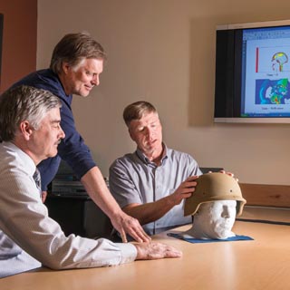
BRAIN POWER — Paul Taylor (5431), right, talks with John Ludwigsen (also 5431), center, and Corey Ford, a neurologist at the University of New Mexico’s Health Sciences Center, about their research on traumatic brain injuries. The three researchers are comparing supercomputer simulations of the physical effects of blast waves on the brain with Ford’s analyses of patients who have suffered such injuries. (Photo by Randy Montoya)
Researchers at Sandia and the University of New Mexico (UNM) are comparing supercomputer simulations of blast waves on the brain with clinical studies of veterans suffering from mild traumatic brain injuries (TBI) to help improve helmet designs.
Paul Taylor and John Ludwigsen of Sandia’s Terminal Ballistics Technology Dept. 5431 and Corey Ford, a neurologist at UNM’s Health Sciences Center, are in the final year of a four-year study of mild TBI funded by the Office of Naval Research. The study is the only TBI research that combines computer modeling and simulation of the physical effects of a blast on the human brain with analyses of clinical magnetic resonance images (MRIs) of patients who suffer such injuries, Paul says.
“Our ultimate goal is to help our military and eventually our civilian population by providing guidance to helmet designers so they can do a better job of protecting against some of these events we are seeing clinically and from a physics perspective,” says Paul, Sandia’s principal investigator on the project. “To do that we’ve got to know what are the threshold conditions that correlate with various levels of TBI.”
Immediately following blast waves, soldiers can be stunned or suffer brief losses of consciousness, but more damage evolves weeks later, Ford says. The symptoms — headaches, memory loss, mood disorders, depression, and cognitive problems — can prevent sufferers from working, he says.
Paul is applying shock wave physics to understand how sensitive brain tissue is affected by waves from roadside bombs or blunt impacts within the first 5-10 milliseconds of exposure. That’s well before a victim’s head moves any significant distance in response to the blast or impact.
“This stuff is over before you have any chance to react and probably before you even knew it happened to you,” Paul says. As teenagers, humans’ fastest reaction times are 75-100 milliseconds.
Ford says levels of energy transmitted into the brain by a blast wave “could be part of the injury mechanism associated with TBI and the mechanism by which it happens may not be mitigated by traditional methods of protecting the head with a helmet.”
At Sandia, the researchers created a computer model of a man’s head and neck. The model includes the jaw — another first in TBI research — because a lot of blasts come from improvised explosive devices (IEDs) at ground level, sending waves traveling at the speed of sound through the jaw and facial structure before they reach the brain, Paul says.
Visible Human Project data used
Sandia’s team used the National Library of Medicine’s Visible Human Project — established in 1989 to create a digital image library of volumetric data representing complete, normal adult male and female anatomy — as a guide to build the head and neck model.
Using images of the male, whose age was close to that of most military personnel, Paul, with Ford as a medical consultant, created geometric models of the seven tissue types in the human head — scalp, bone, white and gray brain matter, membranes, cerebral spinal fluid, and air spaces. Over a year, they catalogued each of the tissue types seen in about 300 “slices” of a cadaver’s head, dividing what they saw into one-millimeter cubes and assigning each a tissue type for the computer simulation.
Paul also imported digitally processed, computed tomography (CT) scans of various helmet designs into the simulations to assess the protective merits of each against blast loading.
In a typical blast simulation, 96 processors on Sandia’s Red Sky supercomputer take about a day to process a millisecond of simulated time, Paul says. Since each blast scenario is simulated out to at least 5 milliseconds, each event can take about a week to calculate.
The 3-D simulations are visualized using two-dimensional multicolored images of a man’s head that record an enormous amount of data. Paul and Ford have focused three types of energy entering the brain that may cause TBI: compressive isotropic energy associated with crushing; tensile isotropic energy that tends to expand parts of the brain and could lead to cavitation; and shear energy that causes distortion and tearing of soft tissue. The pressure and stress within the brain show up on videos created from simulation colors moving in slow motion through and around the brain cavity.
On the clinical side, Ford studied 13 subjects who suffered mild TBI after IEDs exploded near them. Some were stunned, most lost consciousness at least briefly, and most cannot hold a job, he says.
The research partners hope to recruit more patients, especially active military personnel or veterans, who were exposed to blasts that did not penetrate the skin and who suffered a loss of consciousness, Ford says. Candidates must have no history of other blunt traumas.
A battery of tests measured the subjects’ memory, language, and intelligence. These results were correlated with changes in functional magnetic resonance imaging (fMRI) from the patients. The 3-D fMRI studies can detect and map networks in the brain used for processes like movement, vision, and attention. By comparing this data with those of a control group, Ford identified a subgroup of networks displaying abnormal brain activity in the patients. These results were then compared with energy deposition maps predicted by the computer simulations.
The research showed that certain regions of patients’ brains are hyperactive, perhaps because they are compensating for adjacent, damaged areas of the brain that were hit with high energy from the blasts. The hyperactive regions were those predicted to experience the least shear and tensile energies, according to the computer simulations, which can be used to predict where the hyperactivity will likely occur, they say.
Validating simulation with clinical reality
The studies also showed problems with how the patients used visual information, which corresponded to their complaints about having difficulty with attention spans, Ford says.
“This is our way to validate what the simulation shows with the clinical reality,” he says.
Once Paul and Ford determine exactly how and where the wave energy deposited in the brain gives rise to injuries, they can identify threshold levels of stress and energy that cause TBI for consideration by helmet designers, Paul says.
Eventually, Paul says these thresholds could be used for sports helmets and even to place sensors on helmets that would show whether a blast was strong enough to have caused TBI. Currently, many TBI sufferers experience no immediate symptoms that would tell them there’s a problem and cause them to seek medical attention. The sensors could be used to alert them to the problem.
“I want us to be able to understand the physical mechanisms that lead to TBI. It would also be useful if we could make the connection between blast loading and blunt impact trauma,” Paul says. “Once we understand that, we can be more comprehensive in how we protect both our warfighters and athletes against these sorts of injuries.”