In the world of cellular biology, the fact that lipid membranes sometimes spontaneously bend is an accepted phenomenon, but no one has ever conclusively explained the mechanisms driving the curvature. In a research project that brings together synthetic chemistry, bioengineering, and advanced microscopy, Jeanne Stachowiak (8125), Carl Hayden (8353), and Darryl Sasaki (8621) think they may have come across a piece of the answer.
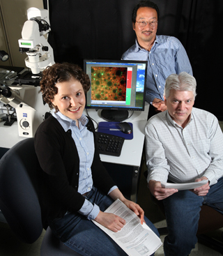
MERGING THEIR INDIVIDUAL AREAS of expertise, Carl Hayden (seated), Jeanne Stachowiak, and Darryl Sasaki discovered that creating domains bound by proteins on vesicles caused the formation of tubules. (Photo by Dino Vournas)
“We discovered that certain confining structures can amplify membrane bending by concentrating the steric interactions between bound proteins,” explains Jeanne. “We took a step back and looked at longer length scales, rather than thinking about a single protein interacting with a single lipid. What we found indicates that when proteins crowd into a small area of the membrane they may be able to cause it to bend.”
Their work was published in a paper, “Steric confinement of proteins on lipid membranes can drive curvature and tubulation,” that appeared in the April 27 issue of the prestigious journal PNAS, the Proceedings of the National Academy of Science.
This insight provides a new way to think about the membrane bending that occurs during basic cellular functions such as endocytosis, the process in which cells absorb external molecules by engulfing them in the cell membrane. Understanding such processes could someday enable programmable biological materials that could be used in far-ranging applications like drug delivery or nanomedicine.
Looking for something else
Like so many scientific discoveries, Jeanne, Carl, and Darryl’s breakthrough came when they were looking for something else. Darryl has been working on lipids with a tunable affinity for proteins for 15 years, since his days as a postdoc at Caltech. He and Carl have collaborated on several projects studying ways to bind proteins to a molecular surface, including a Laboratory Directed Research and Development project called “Biomolecular Transport and Separation in Nanotubular Networks.”
Jeanne returned to Sandia in August 2008 after finishing her PhD at the University of California, Berkeley, in Dan Fletcher’s lab, where her research focused on “giant,” that is, cell-sized, vesicles with a single lipid layer.
“Cell-sized vesicles are useful for imaging,” explains Jeanne. “For fun, Darryl wanted to study his protein-binding lipids in a cell-sized vesicle.”
When Darryl and Carl previously imaged those membranes on glass surfaces, the protein-rich domains that formed were stable micrometer-sized structures. Why they did not coalesce into larger structures was hypothesized to be due to interaction with the substrate. Sure enough, on a giant vesicle the protein-rich areas joined together to form one large domain.
“We wanted an image of protein localized to that domain. Surprisingly, when we added protein we saw these long tubes forming,” says Jeanne.
Initially the researchers thought they had stumbled on an interesting self-assembly concept. For further study, Jeanne went back to Fletcher’s lab at Berkeley to use the fast-imaging confocal microscope. By talking with postdocs in the lab, Jeanne realized that they could be looking at a previously undescribed mechanism.
Getting the right people together
“It was really one of those situations of getting the right group of people together. One of my colleagues at Berkeley who studies endocytosis gave me some papers to read and we suddenly realized that the mechanism we observed could be relevant to cellular processes,” she says. “To us it seemed like a materials phenomenon because we had engineered a synthetic interaction between the protein and the lipid. I wasn’t aware of the significance of membrane bending. It was exciting to learn that these confining structures exist in cells and, in the areas where they may be important, there is not a full explanation for the extreme membrane curvature.”
Jeanne, Darryl, and Carl presented their work at several conferences this spring and were pleased with the positive response. They were also pleased when they learned that Harvard professor George Whitesides, founder of 12 companies including Genzyme and one of the founders of the self-assembly field, served as the editor of their paper.
The work is funded in part by DOE’s Basic Energy Sciences Program in materials science and chemical sciences, which supports the development of biological concepts to create next-generation materials and new approaches to imaging molecular/nanoscale assemblies and fast chemical processes. Jeanne explains that the kind of dramatic reorganization they demonstrated in their paper could be useful as a basic concept for building soft programmable materials.
Now, the researchers are employing microfluidic methods to control the networks of tubules and vesicles that randomly form with the high-affinity protein sites. Such control would represent a significant step toward creating programmable biological materials that could be used in applications like picoliter-scale fluidic transport systems, drug delivery systems, and reconfigurable nanocomposite architectures for sensing, separations, catalysis, and optics.