Under the right conditions, nanotubes may form between human cells with surprising ease, Sandia researchers have found.
The tunnel-like structures have been recognized only in the past few years as tiny but important bodily channels for the good, the bad, and the informational.
“Lipid nanotubes provide a vast array of functions that could greatly impact our approach to treating infection and in understanding how cells respond to pathogens,” says Darryl Sasaki (8331) of Sandia’s Bioscience and Energy Center.
“Our work is the first to show that the formation of nanotubes is not complicated, but can be a general effect of protein-membrane interactions alone.”
Understanding how cellular nanotubes form has become important to medical science over the past few years because of the discovery of what they transport. They seem to serve as routes that protect retroviruses and bacteria as they pass from diseased cells to healthy ones — a fact that may explain why vaccines do poorly against certain invaders. Conversely, the nanotunnels also seem to help trundle bacteria to their doom in the tentacles of microphages. Lastly, the nanotubes may also provide avenues for cells to send and receive information (in the form of chemical molecules) from cell to cell far faster than their random dispersal into the bloodstream would permit.
Given the discovery of this radically different transportation system operating within human tissues, it was natural for researchers to attempt to duplicate the formation of the nanotubes. In their labs, they experimented with giant lipid vesicules that appeared to mimic key aspects of the cellular membrane.
Giant lipid vesicules resemble micron-sized spherical soap bubbles, with the inner side hydrophobic and the outer side hydrophilic.
The object for experimenters was to create conditions in which the spheres would morph into cylinders of nanometer radii.
But researchers had difficulties, says Darryl, perhaps because they used a composite lipid called egg PC that requires unnecessarily high energies to bend into a tubular shape.
Egg PC is inexpensive, readily available, and offers good, stable membrane properties. It is the usual lipid of choice in forming nanocylinders via mechanical stretching techniques.
But Sandia postdoctoral student Haiqing Lui (8331) instead used POPC — a single pure lipid requiring half the bending energy of egg PC.
She was trying to generate nanotubes by a completely different approach that involved the use of motor proteins to stretch naturally occurring membranes into tubes.
Working with George Bachand (8331), she serendipitously found that interaction of the POPC membrane with a high-affinity protein called streptavidin alone was enough to form the nanotubes.
“Perhaps this information — linking membrane-bending energy with nanotube formation — may provide some clue about the membrane structure and the cell’s ability to form such intercellular connections,” Darryl says.
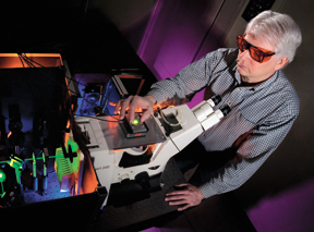
The formation was confirmed by Carl Hayden (8353), who characterized the nanotube formation through a confocal imaging microscope. The custom instrument allows pixel-by-pixel examination of the protein interaction with the membranes comprising the nanotubes by detecting the spectrum and lifetimes of fluorescent labels on the proteins.
Nanotube formation had been noticed previously by cell biologists, but they had dismissed the tiny outgrowths as “junk — an aberration of cells growing in culture,” says Darryl. “The reason they were only noticed recently as trafficking routes is because of labeling studies that marked organelles and proteins. This allowed a focused look at what these nanostructures might be used for.”
It became clear, says Darryl, that the organelles were being transported with “specific directionality” on the backs of motor proteins within the tubes, rather than randomly.
Three-dimensional networks of nanotubes also are found to be created by macrophages — part of the police force of the body — grown in culture, says George. The tubes in appearance and function resemble a kind of spider web, capturing bacteria and transporting them to the macrophages, which eat them.
Other paper authors include postdoc Hahkjoon Kim (8353) and summer intern Elsa Abate (8331).
The lipid work is supported by Sandia’s Laboratory Directed Research and Development office. Motor protein work is supported by DOE’s Office of Basic Energy Sciences.
Results were published in the American Chemical Society’s Langmuir journal in mid-March.