Certain fish species blend with their environment by changing color like chameleons. Their tiny motor proteins carry skin pigment crystals in their “tails” as they walk with their “feet” along the microtubule skeletons of cells to rearrange the animal’s color display.
In two recent papers, Sandia researchers have demonstrated that, in theory, they could produce a similar color change to enable synthetic or hybrid materials to change color like fish do.
“Military camouflage outfits that blend with a variety of environments without need of an outside power source — blue, say, when at sea, and then brown in a desert environment — is where this work could eventually lead,” says principal investigator George Bachand (1132). “Or the same effect could be used in fabricating chic civilian clothing that automatically changes color to fit different visual settings.”
The power source for both the biological and the lab method relies on the basic cellular fuel called ATP, which releases energy as it breaks down. Fifty percent (roughly) is absorbed by the motor proteins.
To switch motor proteins on and off, nature uses complex signaling networks. The Bachand group’s switch is simpler. It involves the genetic insertion of a new binding pocket — a kind of docking port — in the motor protein’s structure. What’s bound and released are zinc ions. Bound zinc ions turn the protein’s action to “off.” Stripping zinc ions out with chemical agents allows the motor protein to work again. The effect is controllable, and even reversible.
Introducing an on/off switch
“We essentially reengineered the protein structure to introduce an on/off switch into the motor,” says George. “So we can now turn our nanofluidic devices on and off.”
Previous efforts at regulating motor activity have used fuel intake as a control mechanism: the less the fuel, the slower the process. The Bachand group’s switch operates independently of fuel by reversibly “freezing” the motor. The advance resembles the improvement in early automobile technologies when a simple ignition switch took over for more complicated rheostats. The paper describing this work was a spotlighted article in the journal Biotechnology and Bioengineering (vol. 100, p. 478).
But what is it that the switch operates?
In a cover article in the high-profile journal Advanced Materials (Dec. 2, 2008), the Sandia team describes a kind of inverted cellular world where motor proteins do not run about but instead are upended so that their tails are embedded in a protein-modified layer on a glass slide. Microtubules — cylindrical protein filaments — instead of forming the stationary cellular skeleton of cells, are passed along by the waving feet of the motor proteins like crowd surfers at a rock concert, or like buckets passed hand-to-hand along a line of firefighters.
Protein-coated quantum dots
Coating the biotin (vitamin H)-modified microtubules are protein (streptavidin)-coated quantum dots — nanoscopic groups of atoms that emit light, their frequency dependent on dot size.
Though the dots operate differently from pigment crystals — the dots do not emit the same frequency of light that they adsorb, while the biological system merely reflects incoming wavelengths — they can perform a similar coloring function.
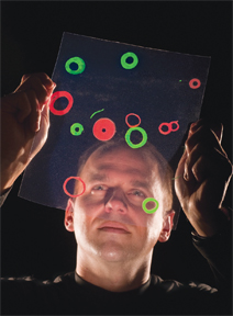
When the motors transport the microtubules and collisions occur, the microtubules tend to stick together and twist until they resemble the cord of a desk phone. The twisting process ultimately forces the formation of stable rings of approximately five micrometers diameter. Their docked quantum dots of cadmium selenide produce a particular range of light frequencies. When mechanical strain in the rings causes them to rupture, the cracked segments are tugged out by the nearby motors until the ring is completely disassembled. The formation and destruction of the two states — free microtubules and rings — can also be reversibly controlled.
A tug-of-war between motor groups
The process resembles the action of fish color changes, which require one group of motor proteins carrying pigments to be “on” all the time while a second group of motor proteins is turned on by complex biological processes at the right time. This produces a tug-of-war between motor groups that results in pigment dispersion and ultimately a color change. When the second motor is switched off, the color returns to the ground aggregate state.
“Our overall process mimics the fish,” says George. “We essentially go from a dispersed particle state to a concentrated one and then back again to dispersed, similar to the fish. Thus, in principle, the mechanism could produce a color change. The underlying science provides a new basis for materials scientists to begin working toward real-world applications.”
The work was supported by DOE Basic Energy Sciences and Sandia’s LDRD office.
Key contributors to the Biotechnology & Bioengineering paper were Adrienne Greene (1132) and Amanda Trent (now a graduate student at University of California, Santa Barbara). Advanced Materials paper contributors were Haiqing Liu (now at Los Alamos National Laboratory), Erik Spoerke (1816), Marlene Bachand (1132), Steve Koch (former Sandia, now an assistant professor at the University of New Mexico), and Bruce Bunker (1816).