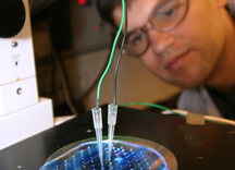
The µChemLab project began with a problem, how to detect trace explosives with a compact, field-portable device. The device relied upon miniaturizing a standard laboratory technique for separating mixtures of components as they move through a column under an electric field — chromatography. But the hair-thin chromatographic columns, shrunk and coiled onto a microfluidic chip, suffered from a “racetrack” effect at the turns, so that particles being separated on the outside of the curves had farther to travel, which smeared the sharp peaks needed for identification.
Unfortunately, the theory behind this transport problem was too complex to model on supercomputers, but Eric Cummings, a principal investigator during the 10-year development funded through Laboratory Directed Research and Development projects, saw that the velocity aspect could be represented by a simple equation with a few constraints, leading to a theory for ideal electrokinetic flow.
Mathematicians Stuart Griffiths (8700) and Robert Nilson (8764), meanwhile, simulated the transport of chemicals in channels in a six-month effort that led to patented turns and bends that minimized dispersion.
Still, Eric believed optimizing any given case should be shorter. “In the back of my mind it seemed there had to be a general solution that was very simple because the theory behind it was so simple,” he says.
He had been trying to verify ideal electrokinesis experimentally. Particles moved differently at boundaries, so the team decided to expand the number of boundaries by creating channels with posts in the middle so they could study behavior there. Posts made the process of moving and sorting particles akin to having racers navigate a forest instead of an open plain — albeit a very small one — since the channels run 50 to 100 microns wide and 5 to 50 microns deep. They also permitted effectively moving particles under an applied voltage that was low enough to not heat and possibly harm the sample.
Working with Anup Singh (8321), Eric investigated using fluorescent liposomes to track the progress of particles through the channels. Their conductance made them behave poorly as markers for electrokinetic mobility. They tended to stream along the post arrays in a phenomenon known as dielectrophoresis, particularly if the applied field were at an angle to the array, and to concentrate in regions over time.
That unforeseen development led to the creation of a sorting and trapping technique known as insulating dielectrophoresis, or iDEP (because the posts are made of a material, such as silica or plastic, that is electrically insulating).
Trapping by tilting the array created non-uniform fields. So overall flow would not be affected, they conducted a quick analysis to find conditions needed to keep the fields uniform within the channel regions.
The analysis indicated that varying the channel depths by interspersing deeper regions with more shallow “spillways” could not only provide uniform fields on each side of the junction, it also allowed designers to incorporate angles in the shallower regions to turn the flow without causing dispersion. This ability to turn the channels permitted creating networks by using calculations simple enough to perform on a calculator.
This breakthrough, several years after the research started, now offered a general solution to the initial dispersion problem.
Experimental investigations of microfluidic channels containing cross-channel ridges designed using Eric’s spatially uniform field approach resulted in two publications in the fall of 2005 in Analytical Chemistry. One demonstrated the influence of manufacturing limitations on fluid flow in ideally designed channels and the other demonstrated continuous separation and concentration of bacterial cells. The work was conducted by postdoctoral researchers Andrew Skulan and Louise Barrett in Microfluidics Dept. 8324 under principal investigator Greg Fiechtner.
Eric’s approach has also led to other devices. One sorts particles into parallel streams by their volume and conductivity and is referred to as a particle spectrometer.
The team has found it useful to design these microfluidic spectrometers with arrays of ridges patterned through photolithography.
“Once you’ve paid for one ridge you can have more,” Eric says. They call this latest approach a corduroy design methodology.
With it, they have concentrated materials by a factor of 6,000 in 16 seconds, although the upper limit is just a function of how much flow can be pushed through. They have also broadened the concept to concentrate, mix, purify,
filter, or sort molecules through perturbing the flow in a variety of ways, besides using an obstacle such as a ridge or valley.
The advantages are that the separations can occur at dramatically higher rates than conventional methods, and they can be based on mechanical or electrical properties not previously exploited by other methods. The speed comes about because separations occur throughout the entire breadth of the microfluidic channel, since they rely on bulk behavior that occurs in a gradient, rather than surface phenomena requiring interaction with a boundary. Theoretically, a large protein might be separated in 10 milliseconds — five orders of magnitude quicker than through conventional chromatography.
A further advantage is that to handle a greater volume, the process could be carried out continuously and in parallel.
The team members believe they are heading toward near-instantaneous separations and manipulations of cells, proteins, and other molecules that can aid research in genetics, proteomics, or sorting and preparation of novel materials; development of medical diagnostic devices; and rapid detection of biological or chemical incidents, among anticipated applications. About such applications Andrew notes, “You can really let your imagination run wild. It’s a very simple design, but there is just a wealth of different behaviors we can obtain by varying the conditions we apply.”