Confocal Raman microscopy and multivariate curve resolution (MCR) can be used to provide molecular information on the microscopy scale, particularly for non-fluorescent biomolecules. Here, we used these techniques to deconfolute spectral signatures for astaxanthin, β-carotene to astaxanthin occurs in lipid vesicles outside of the chloroplast. |
|
Genetically modified cyanobacteria are potential biocatalysts for fuel production. Unlike eukaryotic algae, cyanobacteria are not natural lipid producers, yet cyanobacteria can be easily manipulated through genetic modification. In this work, the metabolism of a model cyanobacterium, Synechococcus elongatus PCC 7942, is engineered for the production of free fatty acids, a precursor for biodiesel production. The engineered strains are characterized using Sandia's hyperspectral fluorescence imaging technology in addition to other traditional techniques to assess the effect of free fatty acid production on the cyanobacterium's physiology and metabolism. |
|
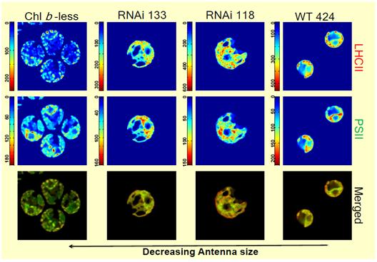
Unicellular green algae harness solar energy using carotenoids, Chl a, and Chl b. We investigated wild-type and mutant lines of Chlamydomonas reinhardtii with truncated light-harvesting antennas to probe how decreasing Chl content influences global architecture of the chloroplast and pigment distribution. MCR analysis of these cell lines yields two spectral components representing LHCII and PSII. |
|
Wild-type cells show typical chloroplast morphology. LHCII and PSII were generally co-located; however, the chloroplast periphery appeared to be enriched in LHCII and the interconnecting regions had more PSII. The Chl b-less mutant was largely devoid of LHCII and the chloroplast morphology had regions that were punctate. The two intermediate-sized antenna mutants (RNAi118 and RNAi133) had spectral component distributions similar to wild-type cells; however, the overall chloroplast morphology was disorganized. In all cell lines, the concentration of PSII is equivalent. |
|
|
|
|
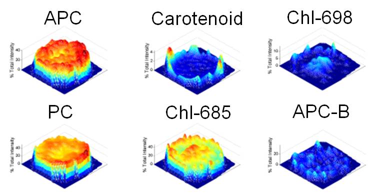
Hyperspectral imaging allows for the identification and quantification of many more fluorescent species. This is especially powerful when dealing with samples that contain photosynthetic pigments, such as those found in plant tissue or those associated with cellulosic and algal biomass energy feedstocks. Sandia and Monsanto are teaming, through a Cooperative Research and Development Agreement (CRADA), to support our joint goal of understanding the photosynthetic properties of plant tissue important for the production of food and biofuel crops. The images to the left illustrate the advantage of using hyperspectral fluorescence imaging compared to the commercially available filter-based microscopy for analysis of crops such as corn leaves. Below are the concentration profiles of a single cyanobacterium (Synechocystis, 2 mm in size) and the associated spectral components. Hyperspectral imaging allowed us to identify six photosynthetic pigments and the spatial location of these pigments at the cellular level. |
||||
|
|
|
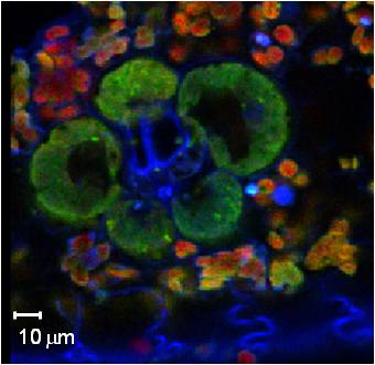
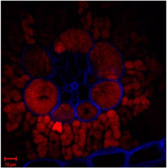
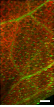
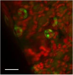
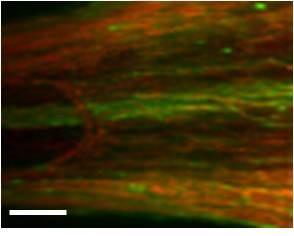
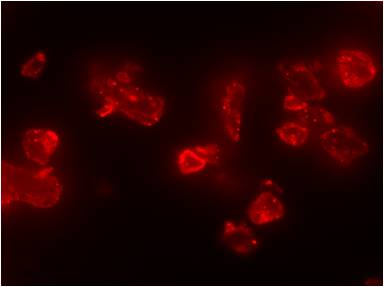
We are using multi-scale hyperspectral fluorescence imaging to visualize enzymes expressing GFP simultaneous with the endogenous chlorophyll fluorescence in the model plant, Arabidopsis thaliana. The images show multivariate curve resolution analysis of multiscale hyperspectral images from A. thaliana (At) expressing green fluorescent protein (GFP). Red: four endogenous (different chlorophyll species); Green: GFP Arabidposis leaf tissue. |
||
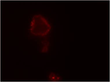
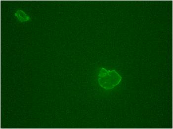
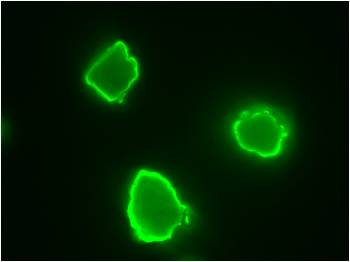
In work with the Joint BioEnergy Institute to optimize the activity of cellulase enzymes on amorphous cellulose, we are characterizing the binding properties of various cellulose binding modules (CBMs). The top two images on the right show that CBM3c binds far more strongly to crystalline cellulose than to amorphous cellulose. The bottom two images show that the addition of CBM3b to glycosyl hydrolase 5 substantially improves binding to amorphous cellulose. This work supports a large enzyme engineering effort to couple CBMs with catalytic domains to design an enzyme cocktail optimized for ionic-liquid treated cellulose. |
|
©1997-2011 Sandia Corporation | Questions and Comments | Privacy and Security | ![]() News release RSS feed
News release RSS feed