Researchers work on new way to image the brain
Sandia researchers want to use small magnetic sensors to image the brain in a way that’s simpler and less expensive than the magnetoencephalography system now used.
Magnetoencephalography is a noninvasive way to measure tiny magnetic fields produced by the brain’s electrical activity. The measurements, able to catch activity as fast as a millisecond, help identify how parts of the brain function and can locate sources of epilepsy and other anomalies.
The state of the art is an array of hundreds of magnetic sensors placed around the head to image the brain by responding to tiny changes in its magnetic fields — sensors called SQUID magnetometers, for superconducting quantum interference device magnetometers. Such systems require magnetic shielding for an entire room and use liquid helium, a cryogen that operates at 4 degrees above absolute zero. Those expensive requirements limit accessibility.
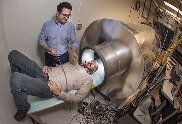
OPM ARRAY — Sandia postdoctoral appointee Amir Borna aids principal investigator Peter Schwindt in entering the person-sized magnetic shield in preparation for a magnetoencephalography measurement with their optically pump magnetometer array. (Photo by Randy Montoya)
Sandia is developing an optically pumped magnetometer, or OPM, sensor array that fits against the head and is housed inside a human-size shield resembling an MRI tube. It avoids the need for cryogenic temperatures or a shielded room, so it would be easier and cheaper to use.
That would make magnetoencephalography more useful for neurology in diagnosing and studying brain conditions and for cognitive science, including emerging research on post-traumatic stress disorder and traumatic brain injury, say the project’s principal investigator Peter Schwindt and former Sandia manager Rob Boye.
“Who’s not interested in brain science?” Peter says. “It’s fascinating stuff.”
The Sandia team published a paper in November in Physics in Medicine and Biology that demonstrates Sandia’s system can detect signals from the brain. The team published a paper last year in Optics Express describing their OPM sensor.
Demo system developed during four-year project
During a four-year project funded by the National Institutes of Health, Sandia built a prototype magnetoencephalography system with the OPM array placed inside a person-size magnetic shield. The OPM is a quantum sensor that includes a small glass cell containing a gas of rubidium atoms, a pump laser to set the state of individual atoms in the gas, and a probe laser to read the changing state of the atoms. Change in state depends on the strength of the brain’s magnetic field sensed by the array.
The demonstration system featured 20 magnetometer channels in five sensors covering less than a quarter of an adult’s skull. The team wants to image more of the brain in the future by developing an array that covers the whole head, like today’s SQUID systems.
“Just because you can detect a magnetic field doesn’t mean you know where it’s coming from.”
Sandia compared its findings to those from a commercial SQUID system, using neurology tests that produce well-understood results. One test sounds a quarter-second-long tone in both ears, producing a spike in the auditory cortex. Another test, a nerve stimulus, causes a thumb twitch, resulting in a response in the somatosensory cortex. Both responses are readily observed with Sandia’s system, and the team uses both responses to characterize and refine its system.
“In essence, you can think of the atoms as little spinning tops,” Rob says. “When there’s a magnetic field present, it’ll make those tops rotate. The probe laser can sense that rotation. In your brain, when a bunch of neurons fire, there’s a little electrical current. Current gives rise to a magnetic field, so it’s the flow of charges in your neurons that gives rise to the magnetic fields sensed by the OPM.”
Commercial SQUID arrays use fixed helmets, with a head-to-sensor distance of at least 2 centimeters and 10 cm or more for children, Peter says. Because Sandia’s array conforms to the head, the head-to-sensor distance is shorter and constant. The team wants to reduce its current distance of 1.2 cm to 0.5-0.7 cm, since the quality of signals from the brain drops off quickly with distance, Peter says.
Making magnetoencephalography more available
Dr. Bruce Fisch, retired professor emeritus at the University of New Mexico Health Sciences Center and former director of UNM’s clinical magnetoencephalography program, says Sandia’s work could make magnetoencephalography more widely available. Fisch, who consulted on the project, says in evaluating epilepsy patients for surgery aimed at stopping seizures, it’s important to locate the source of brain signals more precisely than possible with the more familiar MRI. UNM uses the SQUID system at the Mind Research Network to perform clinical magnetoencephalography scans, Fisch says.
Peter says it’s too early to estimate how much an OPM-based system would cost. Depending on factors such as auxiliary devices, a comprehensive SQUID-based magnetoencephalography system can cost from $1.8 million to $4 million, including a magnetically shielded room, says Miikka Putaala, director of business line magnetoencephalography for Elekta Neuroscience of Finland, which makes such systems.
The next step is to show the system can not only detect signals from the brain, but also pinpoint where the signals originate. Actions such as thinking or contracting a muscle create magnetic fields in the brain, but they’re difficult to isolate.
“Who’s not interested in brain science? It’s fascinating stuff.”
“Just because you can detect a magnetic field doesn’t mean you know where it’s coming from,” Rob says.
The OPM array is placed over different parts of the head to focus the array on specific areas of the brain. Operators combine information to localize the source of the magnetic field to find where the brain is active.
Sandia’s team is using the measured signals to localize sources in the brain. The team is working to improve the imperfect calibration of sensors and knowledge of the OPM array relative to the position of the brain to continue to improve the accuracy of localizing brain activity.
Fitting the array more closely to the scalp can improve localization accuracy and distinguish between closely spaced neuronal sources. A better fitting array also might detect activity that can’t be sensed now.
“In particular, this can be very interesting for pediatric and infant studies of brain development,” Peter says. “The closer you get, the more spatial fidelity you’ll have.”