It all starts with the breach of a single cell. Viruses, bacteria, and other maladies-in-waiting skulk around the body, waiting for a chance to strike. Once they slip past the immune system’s defenses, they start ferociously multiplying, preparing to unleash havoc on an unsuspecting host.
Exactly how and why the cell’s defenses fail against some invaders while successfully fending off others has long been a mystery. A novel super-resolution microscopy technique developed at Sandia is providing new answers to old questions by unveiling never-before-seen detail of the cell membrane. The insights gleaned from the research could open doors to new diagnostics, prevention, and treatment techniques. The research supports Sandia’s biological threat reduction programs and could be expanded to support biofuels research.
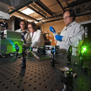
“We’re trying to do molecular biology with a microscope, but to do that, we must be able to look at things on a molecular scale,” says Jesse Aaron (8622), a postdoctoral appointee.
The cell membrane is a bustling hub of activity on a miniscule scale. While providing structure and housing the cell’s interior, the membrane regulates movement of materials in and out of the cell, controls adhesion to other objects, and coordinates the cell’s communications and subsequent actions through signaling. Receptor proteins on the surface of immune cells, known as Toll-like receptors (TLRs), are tasked with recognizing intruders, or antigens. The TLR4 member of this receptor family responds to certain types of bacteria by detecting lipopolysaccharides (LPS) present on their surface. They subsequently initiate signaling to alert the cell and activate an immune response.
A vexing problem
Using imaging techniques they developed, Jesse, Jeri Timlin (8622), and Bryan Carson (8622) discovered that TLR4 proteins cluster in the membrane when confronted with LPS derived from E.coli, which increases cell signaling and response. Interestingly, LPS derived from the bacteria that causes plague, Yersinia pestis, do not cause the same effects. This finding, which marked the first time such small events could be imaged and compared, could explain why some pathogens are able to thwart the human immune system.
Being able to image the cell surface with high enough resolution to see the earliest binding events has been a vexing problem, since even the most sophisticated optical microscopes are bound by the diffraction barrier, which limits what can be resolved using visible light.
“With more traditional visualization methods, you can’t see the level of detail you need. It’s important to look at not only what’s present, but also when and where it’s present in the cell,” says Jeri.
Dual color capabilities
The technique used by Jeri and Jesse builds on superresolution capabilities developed in recent years, but goes another step by adding dual-color capabilities to the relatively new stochastic optical reconstruction microscopy, or STORM. The combination enables the Sandia team to get an exponentially better picture by simultaneously imaging LPS and TLR4 receptors on the membrane.
“Current capabilities are akin to looking out the window of an airplane and seeing the irrigation circles. You know that plants are there, but you can’t tell what kinds of plants they are or what shape the leaves are,” says Bryan, a Sandia immunologist who was an integral part of the project. “But with this technology, it’s like zooming in and seeing the leaves and the structure of the plants. That buys you a lot in terms of understanding the mechanism.”
The NIH awarded Jeri a five-year, $300,000 per year grant in 2009 to develop such visualization power, and it has exciting potential for future applications. Next on the agenda is developing the capability to image live cells in real time using spectral stimulated emission depletion, or STED, technology.
“We’re working toward using a version of superresolution that’s much more live-cell friendly, and extend that in terms of what colors are available to do multiple colors, but still maintain the live-cell friendliness. I see this as a beginning of a long development in this type of imaging technology,” Jeri says.
The eventual goals will likely expand as the technology reveals additional surprising capabilities. Eventually, the Sandia team would like to be able to visualize protein/protein interactions.
Seeing the whole biological process
“Every biological process that goes on in your body is somehow controlled by proteins forming complexes with other proteins or complexes in the membrane, so this would give you this ability to look, with high spatial resolution and multiplexed color capabilities, at four or more things in a living cell, which can’t be done very easily right now. It can be done in pieces, but we want to see the whole biological process,” Jeri says.
The technology has exciting potential in immunology and drug discovery as well, Bryan says.
“We’re hoping to do something like label the viral particles and watch them in real time, or as close as we can to real time, in the internalization process,” he says. “With the super-resolution technique, we can actually watch them move through the membrane and see if there are other structures being recruited by the virus to the site of internalization. That will hopefully give us mechanistic insight into how a given virus enters a cell. Understanding that mechanism can lend itself to identifying drugs or other compounds that might block viral entry.”
The team is interested in expanding the technology’s capabilities to research other areas such as biofuels to better understand where and when different pigments are located on the membrane of oil-producing algae. This would provide valuable insight into their photosynthesis functions, which could help in more efficient biofuel production.
“A lot of this work is still pretty initial, but we’re encouraged by what we’re seeing and excited for future potential,” Jesse says. – Stephanie Hobby