If you’re physically attacked, you hope you’ll respond.
That hoped-for response to threat includes the molecular level: We want our cells to respond defensively when an antigen lands on a cell membrane and prepares to cause mischief.
But to activate a response, a cell must become aware of the presence of the intruder on its membrane, just as a human first must become aware of a mosquito on a forearm in order to slap it.
In a triumph of joint experimental work, physicists at Sandia and biologists at the University of New Mexico’s Cancer Research and Treatment Center have combined techniques amazing to the layperson to make real-time movies that show exactly how a 50-nm-thick cell membrane notifies the cell it encloses that a hostile alien presence — an antigen — has made a landing.
Characterization in real time
“We were able to characterize the motion of the receptor proteins in the membrane in real time as they respond to the antigen,” says lead Sandia researcher Alan Burns (now 1012). “Perhaps more importantly, we learned the cell membrane is really complicated and highly structured, rather than fluid and unstructured, as is the prevailing notion.”
This new information explains why membrane proteins may not always notify the cell nuclei of problems.
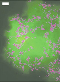
The membrane structures, which resemble holding corrals, says Alan, move around in the membrane. But they restrict the motion of proteins. The response of the cell requires that the antigen receptor proteins cluster with other proteins to commence the cellular signaling network.
“The proteins are like Paul Revere giving a warning,” says Alan. “When proteins bind antigens, they begin to cluster. This causes other proteins to thrash around. That may send a message from the membrane to the cell nucleus that something’s wrong.
“But if there are places on the membrane that are walled off and an antigen lands there, the cell isn’t notified of the problem. No protein, no warning.”
UNM researchers already knew that incoming antigens were detected by proteins present in the lipid matrix of the cell membrane. But how exactly to determine the process?
Alan, working with his former postdoc Keith Lidke (now a UNM professor), modified a special microscope called a total internal reflection fluorescence (TIRF) microscope whose laser-light output is completely contained within the microscope coverslip. This resembles the way optical fibers transport light, except that the TIRF does not ever release any light. But though the light is contained, making its use seem at first an exercise in futility because it penetrates nothing external, it generates a tiny electrical exploratory field that extends about 100 nm into the cell, which lies supported by the coverslip.
“When a cell settles on a piece of bare thin glass,” says Alan, “the membrane of the cell by definition is snuggled up against the glass and available to the radiation field.”
Watching bank robbers
Enter the UNM biology team. Led by professor Diane Lidke (Keith’s wife), the team was able to attach quantum dots of 30 nm and 40 nm respectively to the antigen receptor proteins in the membrane.
Quantum dots emit light when stimulated by an electrical field. The color fluoresced is determined by the size of the dot. So one protein, when stimulated by the laser’s electrical field, emitted orange light. The other emitted red. That way researchers could keep track of the motion of single, individual proteins and see how they interacted; moreover, it allowed them to observe barriers to the motion.
Sensitive CCD cameras picked up and videotaped the motion of the lit-up proteins as they reacted to the introduction of antigens to the membrane.
“It was like using cameras to watch individual bank robbers move around as a holdup progressed,” says Alan.
The work is of interest to Sandia, a national defense lab interested in determining the human response to bioinfectious diseases, and to UNM’s bioscience program.
Other authors on the paper were graduate student Nicholas Andrews and biology professors Bridget Wilson and Janet Oliver, all with UNM’s Cancer Research and Treatment Center.
The Sandia work was funded by its Laboratory Directed Research and Development (LDRD) office. The UNM work was funded by the National Institutes of Health. The work was published online the week of July 20 in the journal Nature Cell Biology.