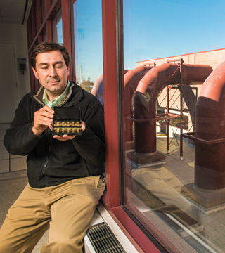
Murat Okandan (1719) holds one of the microscale actuators that could lead to better understanding of brain function, which could help with prevention, diagnostic, and treatment techniques for brain disorders. (Photo by Randy Montoya)
They are with us every moment of every day, controlling every action we make, from the breath we breathe to the words we speak, and yet, there is still a lot we don’t know about the cells that make up our nervous systems. When things go awry and nerve cells don’t communicate as they should, the consequences can be devastating. Speech can be slurred, muscles stop working on command, and memories can be lost forever.
Better understanding of brain function could lead to new prevention, diagnostic, and treatment techniques, but the brain is complex and difficult to study. If you were to hold it in your hand, you would likely marvel at how much your brain feels and moves like Jell-O. This tissue is laced with neurons with tiny cell bodies, which generate electrical signals to control nervous system functions. Those signals can be recorded and measured if a suitably small electrode is in the vicinity, but that presents challenges. Brain tissue is always moving to different degrees in response to the subject’s movement and breathing patterns. In addition, the nerve tissue is incredibly sensitive, and if disrupted by a foreign body, the cells trigger an immune response to encapsulate the intruding probe and barricade it from the electrical signal it’s trying to capture and understand.
Working to develop intelligent neural interfaces
That challenge led Jit Muthuswamy, an associate professor of biomedical engineering at Arizona State University, Tempe (ASU), to pursue a robotic electrode system that would seek and maintain contact with neurons of interest in a subject going through normal behavioral routines. “We are working to develop chronic, reliable, intelligent neural interfaces that will communicate with single neurons in a variety of applications, some of which are emerging and others that are almost to market,” Muthuswamy says. “Things like brain prosthesis are critically dependent on us being able to interface with single neurons reliably over the course of a patient’s life with a prosthetic application.”
Key to the success of the above robotic approach are the microscale actuators that would be needed to reposition the electrodes. This led Muthuswamy in 2000 to seek out Murat Okandan (1719) and the unique microsystems engineering capabilities available at Sandia’s MESA facility.
“The process flow we use to make these isn’t available anywhere else in the world, so the level of complexity and mechanical design space we had to design and fabricate these was immensely larger than what other researchers might have,” Murat says, adding that he has been working with Muthuswamy’s research team since that initial contact to find a suitable method to track individual neurons as they fire.
In the past, probes were made of a sharpened metal wire, inserted in the tissue. The closer the probe is to the neuron, the stronger the signal, so experimenters ideally try to get as close as possible without disrupting surrounding tissue. The problem is that even a thin wire is too big; such a probe can take measurements around the neuron, but is far too cumbersome to be reliable for long durations.
Equally important is capturing the signals from an awake animal; given their size and rigidity, current probes are generally not suited to gather recordings as the animal responds to its environment. Those units are not self-contained, hindering the ability of the animals to move around freely.
The microscale actuators and microelectrode are critical to addressing both of those issues and interacting with individual nerve cells with minimal damage to surrounding tissue. The microscale actuators and associated packaging system developed at ASU and Sandia enable the probe to move autonomously in and out of the areas surrounding the cell collecting measurements while compensating for any movement in the neuron or brain tissue.
About the size of a thumbnail, the self-contained unit has three microelectrodes and associated micro actuators. When a current runs through the thermal actuator, it expands, and pushes the microelectrodes outward over the edge of the unit, which is flat to fit against the tissue. Because the actuator is so small, it can be heated to several hundred degrees Celsius and cooled again 1,000 times per second. It takes 540 cycles to fully extend the probe, but that can be done quickly — in a second or less.
Scale of this system is unique
Thermal actuators have been used for years at Sandia and elsewhere, but the scale of this system is unique. “The idea that we could build this system to achieve multiple millimeters of total displacement out of a micron-scaled device was a significant milestone,” says Michael Baker (1719), who designed the actuator. “We used electrostatic actuators in the past, but the thermal actuator provides much higher force, which is needed to move the probe in tissue.”
The microelectrodes are made of highly doped polysilicon, which the team discovered has a number of advantages. It is almost metal-like in its conductivity, but durable enough for millions of cycles and provides a high signal-to-noise ratio, which is much greater than previous wire probes, and provides high-quality measurement signals.
Muthuswamy and Murat are currently developing the capability to produce richer data with resolution in the sub-micron range to be able to go inside cells and take measurements there. They are also working on stacking the existing chips and decreasing the space between probes. Muthuswamy’s Neural Microsystems lab at ASU has developed a unique stacking approach for creating a three-dimensional array of actuated microelectrodes. “By building a three-dimensional array, we would have access to significantly more information, rather than just a slice,” Murat says. “We’re very encouraged by the progress we have made, and are looking forward to building on that progress.”