Science rarely follows a perfectly charted course. A map of the evolution of scientific discoveries would look like the tributaries of the Amazon River, with one idea leading to another, sometimes extending in a single direction, dead ending, or looping back to the source.
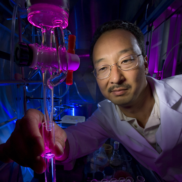
So it went for Darryl Sasaki (8651) and Carl Hayden (8353), when a chemical mixture that didn’t organize quite right enabled them to stumble upon a synthetic analog of an important cellular process. They had been studying protein dynamics, specifically ways to bind proteins to a molecular surface, and discovered a method of creating reversible microdomains that draw proteins onto the surface of the cell like a powerful vacuum. The work recently appeared in the Journal of the American Chemical Society.
The original project headed by Michael Kent (8622) sought to understand how specific protein receptors organize on a cellular surface in response to pathogen presence, which activates our innate immune system. That project, funded by Sandia’s Laboratory Directed Research and Development program, was part of a Sandia strategy in the area of biodefense and emerging infectious disease to elucidate the molecular bases of host-pathogen response in the innate immune system. Previous experiments with other cell membrane models had demonstrated the ability to bind proteins to specific molecules in the membrane while maintaining mobility and dispersion across the surface.
In this case, when the researchers added copper to make the surface adhesive for proteins tagged with a histidine (His) tail, the copper binding molecules formed stable microdomains. In genetic research, a His-tag is frequently used as a way to purify recombinant proteins. The discovery came about because the copper binding molecules changed their electrostatic nature in an unexpected way.
Darryl says they noticed that “instead of becoming positively charged as we had seen with similar systems in the past, the copper binding molecules became charge neutral, allowing the molecules to phase separate from the rest of the membrane.”
Initially the researchers didn’t understand the nature of the dark domains on the surface. First, they thought they had generated holes in bilayer, but through Carl’s experiments to characterize the results, soon it became apparent that the dark domains were formed by the aggregated molecules. Using fluorescent-labeled His-tag proteins they demonstrated that the dark patches were protein-targeted, copper-rich domains.
When they removed the copper, the proteins quickly dispersed. That’s when Carl and Darryl realized they were onto something. In a broad sense, their system mimics the formation and function of membrane microdomains in cells, which are also called lipid rafts.
A controversial concept
“Like lipid rafts, our microdomains are directed to form through a chemical signal and disappear upon removal of the signal, and they perform as sites with enhanced recognition properties for specific agents, such as signaling molecules, proteins, and viral particles,” says Darryl. “Lipid rafts are still a somewhat controversial concept because they have not been observed in live cells, but can be formed in model systems. Their existence could help explain many biological functions, including those related to the response of a host cell to an invading pathogen.”
The work draws upon both researchers’ areas of expertise: Darryl, an organic synthetic chemist, created the molecules and lipid membranes that Carl, a laser spectroscopist, visualized. “This is an example of how Sandia brings together scientists with different expertise and skills to tackle complex problems,” says Darryl. “If we were at a university, Carl would be sitting in a different building and we might not ever cross paths.”
The original project examined protein organization, not the details of membrane structure. But with the ability to toggle protein affinity and nanoarchitecture, the work now impacts Basic Energy Science (BES) research on switchable materials. For Carl’s BES work studying single molecule conformations and function, the ability to switch on or off the binding of molecules to membranes provides new ways to look at how interactions with membranes control functions of biological molecules.
“For BES programs we are trying to develop suitable materials to build unique structures at the nanoscale that can be disassembled easily and quickly,” says Darryl. “The potential for switchable nanoscale materials is huge. Applications might include sensing, light capture, and even nanowires to put function into composite structures.”
Carl and Darryl, in collaboration with Jeanne Stachowiak (8125), are also looking at binding of proteins to the same membrane structures in vesicles. “At certain lipid compositions, the proteins cause the membrane to pucker and generate shapes that look like eukaryotic events occurring. Eventually the structures shrink down to a tube that can grow pretty long. It’s an amazing way to generate even higher order structures,” says Darryl.
Down the road, switchable materials could be used to create nanostructures for picoliter fluidics, addressable nanoparticle coatings, and targeted drug delivery. Switchable materials may even someday be used to create bio-based electronics that assemble and reassemble themselves in response to specific stimuli, leading to materials for numerous applications, including bionic devices.