Modeling, simulations aim to boost understanding of injuries, body armor
Sandia is developing specialized computer modeling and simulation methods to better understand how blasts on a battlefield could lead to traumatic brain injury and injuries to vital organs, like the heart and lungs.
Researchers at the Labs have studied the mechanisms behind traumatic brain injury for about a decade. Their traumatic injury modeling and simulation project began with a head-and-neck representation, and now they’ve created a high-fidelity, digital model of a man from the waist up to study the minute mechanisms behind trauma.
“We’re also concerned about the possibility of injury to the life-support systems in the torso. Everything’s interconnected,” says Paul Taylor, who leads the project. “Clearly, we would love to have a representation of a full human but certainly capturing all the regions where life-critical organs are located is a very good start.”
The information could help manufacturers develop better designs for helmets and body armor.
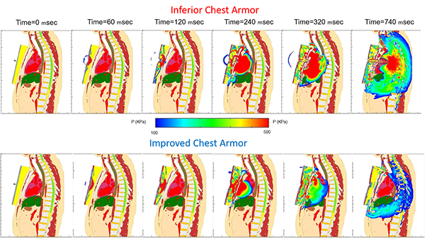
HUMAN MODELING AND SIMULATION — Sandia researchers illustrate their ability to use human modeling and simulation work to compare two notional chest armor designs varying in thickness and material properties. The top row shows a projectile hitting the inferior chest armor design, which failed to keep the projectile from penetrating the chest of the torso model. The bottom row illustrates an improved design. The images show pressure being transferred from the projectile into the torso and moving through the internal organs, which could lead to behind-armor blunt trauma inj
“Protection of the soldier, sailor, airman, or marine is essential, and well aligned with our national security mission against challenging and new lethal threats,” says program manager Doug Dederman. “It is a privilege for our integrated military systems staff to team with the Department of Defense and medical communities to improve both diagnostic capabilities and mitigation of risk with improved protective equipment.”
Sandia’s most recent work grew from a Laboratory Directed Research and Development-funded project that wrapped up in late 2016. Along the way, the team conducted both macroscale and microscale traumatic brain injury simulations, began working with doctors to correlate simulation predictions with clinical assessments of people with brain injury, and increased the size of their team.
They theorize that a phenomenon called fluid cavitation can lead to traumatic brain injury. They’ve developed macroscale simulations to test the hypothesis and extended their work into microscale studies to examine whether blast and short-pulse blunt impact, such as a projectile hitting body armor, could lead to fluid cavitation, forming bubbles whose collapse could damage sensitive brain and lung tissue, Paul says.
Localized shock waves
Cavitation is the formation of vapor cavities — bubbles — caused by rapid pressure changes in fluid, which can occur from blast exposure. Bubbles form and, because they’re unstable, immediately collapse, generating a microjet or miniature localized shock wave. It’s a physics phenomenon commonly seen at the leading edge of spinning ship propellers, eroding those propellers.
“We’ve been able to demonstrate, at least theoretically, that the individual experiences fluid cavitation in the brain"
“We’ve been able to demonstrate, at least theoretically, that the individual experiences fluid cavitation in the brain. We’ve subjected our head-neck model to blast from the front, from the side, from the rear, and what we see are what looks like peppered regions in the brain,” localized regions experiencing cavitation, Paul says, pointing to the occipital, temporal, and brain stem areas on a slide from a simulation.
“Does cavitation occur, and if so, where might it be occurring?” says team member Candice Cooper, who developed the macroscale simulation. “Then we look at those areas on the microscale to see if cavitation is indeed occurring, how might it damage these tissues and lead to traumatic brain injury.”
The smallest area in the macroscale simulation is 1 cubic millimeter, which isn’t small enough to capture the physics of fluid cavitation very well, Paul says.
White matter axons
Enter Shivonne Haniff, who performs microscale modeling and simulation to complement Candice’s macroscale work, simulating the formation and collapse of cavitation bubbles in the brain in scales below 1 millimeter.
One of Shivonne’s models represents axonal fiber bundle tracks within the brain’s white matter. Typically, white matter axons have myelin sheathing, a protective coating, similar to how insulation protects electric wiring. Myelin sheathing accelerates neurological pulses, allowing humans to process information very quickly. Diseases, such as multiple sclerosis, degrade myelin sheathing and drastically reduce pulse transmission.
The team hypothesizes that blast- and impact-induced cavitation and subsequent bubble collapse also could damage myelin sheathing.
Shivonne’s video of a microscale simulation of cavitation bubble collapse within the white matter axon fiber bundle introduces a pressure pulse from one side, causing asymmetric collapse of the bubbles, generating highly localized pressure pulses and microjetting that damages neighboring axons and their myelin sheathing.
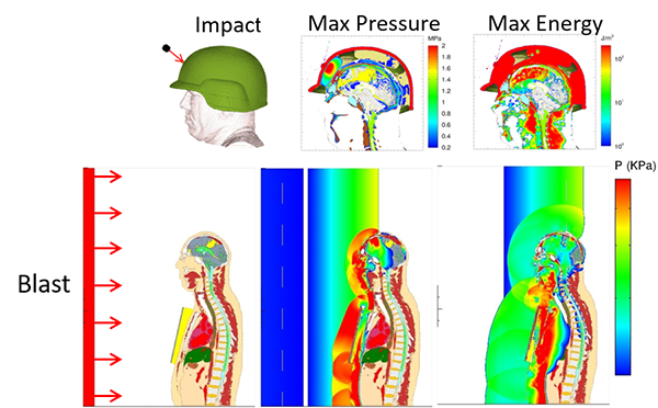
BALLISTIC IMPACT — These images show the implementation of Sandia’s head-neck model (top) and the head-neck-torso model (bottom) in a ballistic impact simulation (top) and a blast impact simulation (bottom). Researchers can measure potential damage variables such as pressure (top center and bottom row), energy (top right), stresses, and strains within the human models to better understand mechanics that could lead to traumatic brain injury or injury to vital organs.
The team studied how compressive wave amplitude and bubble size influenced microjetting strength.
“To assess damage potential from bubble collapse-induced microjetting, we looked at pressures and shear stresses downstream of the bubbles. The shear stresses in the myelin sheathing were considerably higher than the shear stresses in the axon core, indicating the myelin acts as a protective barrier,” Shivonne says. “However, damage to this myelin sheathing could impair the transmission of nerve signals, which can lead to neurological problems.”
"Damage to this myelin sheathing could impair the transmission of nerve signals, which can lead to neurological problems.
She is focusing now on modeling cavitation damage within the blood-brain barrier, a semi-permeable vascular system that allows passage of nutrients and gases needed by the brain but blocks harmful toxins. A video simulation shows cavitation bubbles suddenly collapsing under pressure, drastically increasing pressure and shear loading on surrounding tissue, which can damage it. Simulations look at the effects of different bubble diameters, bubble density, and pressure wave amplitudes on the degree of damage.
Modeling damage mechanisms
Candice also conducted modeling and simulations for a generic body armor configuration. The work was aimed at understanding the modeling problem rather than reaching conclusions applicable to specific armor. Her simulation studied pressures within the heart, lungs, and other organs in different scenarios, such as a soldier standing about 10 feet from a roadside bomb blast.
“We looked at pressure as well as the shearing stress that can lead to tissue tearing, and found that in this notional case, having padding behind the armor actually increased peak pressures in life-critical organs, the heart and the liver, which could lead to damage,” Candice says. “It also led to an increase in shear stresses in all of the organs that we looked at.
“This is just an example of how we can use our modeling and simulation tools. If someone came to us with their armor design and said, ‘Would you take a look at this,’ we could vary the materials of the foam padding, the positioning of the foam padding, the size or geometry of the foam padding or of the armor plate itself,” she says. “We could look at variations on their design and let them know this change makes it better, that change makes it worse.”
The project has a long-term association with Dr. Corey Ford at the University of New Mexico Health Sciences Center and a more recent one with the Air Force San Antonio Military Medical Center. Candice, Paul, and team member Chad Hovey recently presented their research at the International Mechanical Engineering Congress & Exposition, work that was funded through the Military Medical Center. Shivonne and Paul gave a presentation at the same conference, outlining microscale cavitation studies, work funded by the US Office of Naval Research by Dr. Tim Bentley, and published a paper on the topic in a recent edition of Shock Waves. The fifth team member, Ryan Terpsma, has assisted in macroscale modeling of behind-helmet blunt trauma resulting from bullet impacts.
The team also works with experimental collaborators at Los Alamos National Laboratory, New Mexico Tech, its Energetic Materials Research and Training Center, and Michigan State University, some of whom perform blast tube experiments on a physical model. The project recently began working with Team Wendy, a company that manufactures military and civilian helmets.